. 2011:2011:379894.
doi: 10.1155/2011/379894. Epub 2010 Sep 26.

Role of calcium in phosphatidylserine externalisation in red blood cells from sickle cell patients

Affiliations

Role of calcium in phosphatidylserine externalisation in red blood cells from sickle cell patients

Erwin Weiss et al. Anemia. 2011.

Abstract

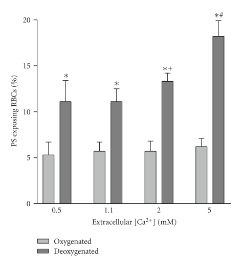
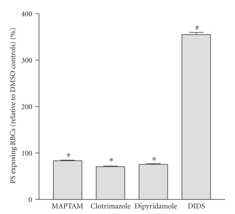
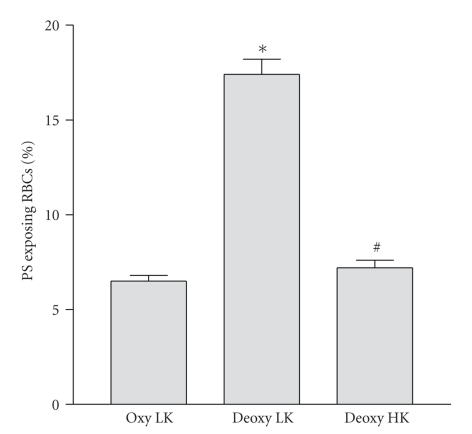
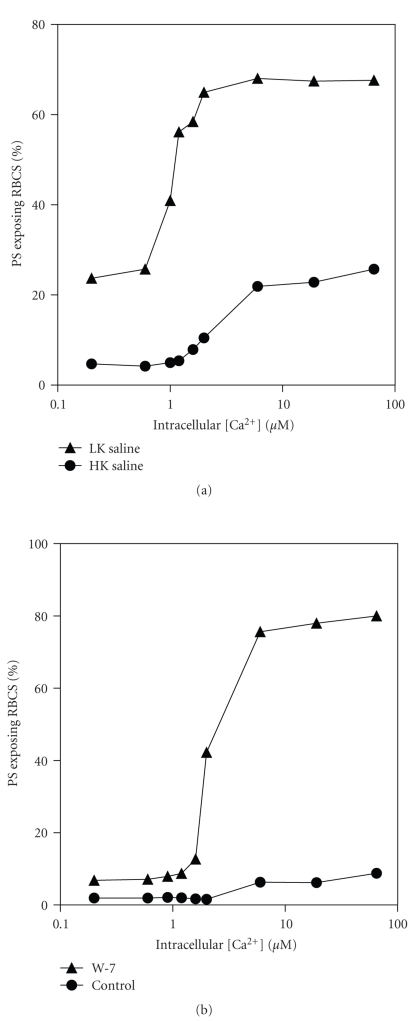
Phosphatidylserine exposure occurs in red blood cells (RBCs) from sickle cell disease (SCD) patients and is increased by deoxygenation. The mechanisms responsible remain unclear. RBCs from SCD patients also have elevated cation permeability, and, in particular, a deoxygenation-induced cation conductance which mediates Ca(2+) entry, providing an obvious link with phosphatidylserine exposure. The role of Ca(2+) was investigated using FITC-labelled annexin. Results confirmed high phosphatidylserine exposure in RBCs from SCD patients increasing upon deoxygenation. When deoxygenated, phosphatidylserine exposure was further elevated as extracellular [Ca(2+)] was increased. This effect was inhibited by dipyridamole, intracellular Ca(2+) chelation, and Gardos channel inhibition. Phosphatidylserine exposure was reduced in high K(+) saline. Ca(2+) levels required to elicit phosphatidylserine exposure were in the low micromolar range. Findings are consistent with Ca(2+) entry through the deoxygenation-induced pathway (P(sickle)), activating the Gardos channel. [Ca(2+)] required for phosphatidylserine scrambling are in the range achievable in vivo.


Figures

Figure 1
Figure 1
Effect of oxygen tension and extracellular Ca2+ on phosphatidylserine (PS) exposure in red blood cells (RBCs) from sickle cell patients. RBCs were incubated for 18 hours at four extracellular [Ca2+]'s (0.5, 1.1, 2.0 and 5.0 mM) after which they were labelled with FITC-annexin (as described in Section 2). Histograms representing mean percentage of positive RBCs ± S.E.M. for 5 different patients. *P < .01 deoxy compare to oxy; + P < .05 cf 0.5 mM Ca2+ deoxy; # P < .01 cf 0.5 mM Ca2+ deoxy.
Figure 2
Figure 2
Effect of inhibitors on phosphatidylserine (PS) exposure in red blood cells (RBCs) from sickle cell patients. RBCs were incubated under deoxygenated conditions (1% O2) for 3 hours (5 mM extracellular [Ca2+]) after which they were labelled with FITC-annexin. Four conditions (all with 0.5% DMSO) are shown: MAPTAM-treated RBCs (loaded with 5 μM MAPTAM prior to deoxygenation), clotrimazole (10 μM), dipyridamole (50 μM), and DIDS (50 μM). Results are presented as percentage PS exposing RBCs relative to control RBCs exposed to 0.5% DMSO only. Histograms represent means ± S.E.M. (n = 3). *P < .01 and # P < .0001 cf DMSO controls.
Figure 3
Figure 3
Effect of extracellular K+ on phosphatidylserine (PS) exposure in red blood cells (RBCs) from sickle cell patients. RBCs were incubated for 18 hours with extracellular [Ca2+] of 5 mM under oxygenated (20% O2) or deoxygenated (1% O2) conditions in either low K+-containing (extracellular [K+] of 5 mM) saline or high K+-containing (90 mM [K+]) saline. Histograms represent means ± S.E.M. (n = 3). *P < .001 compare to LK oxy; #N.S. cf. LK oxy.
Figure 4
Figure 4
Effect of manipulation of intracellular Ca2+ on phosphatidylserine (PS) exposure in red blood cells (RBCs) from sickle cell patients. RBCs were first treated with vanadate (1 mM) to inhibit the plasma membrane Ca2+ pump and also the aminophospholipid translocase (flippase) before addition of bromo-A23187 (1.2 μM, 1% haematocrit) and requisite extracellular [Ca2+]s for 30 min. They were then treated with Co2+ (0.4 mM) before labelling with FITC-annexin. Intracellular [Ca2+] is calculated from extracellular [Ca2+] × r 2, where r 2 was taken as 2.05 [36]. Results presented are from a single experiment representative of 5 others.
Figure 5
Figure 5
Ca2+ loading of red blood cells (RBCs) from sickle cell patients. RBCs were loaded with the Ca2+ fluorophore fluo-4 (see Methods). They were then incubated for 30 min in the absence (left—thin line) or presence (right—thick line) of bromo-A23187 at an extracellular [Ca2+] of 1 μM. Results are presented as histogram of fluorescence of a single experiment representative of 3.
Figure 6
Figure 6
Effect of K+ and calmodulin inhibition on Ca2+-induced exposure of phosphatidylserine (PS) in red blood cells (RBCs) from sickle cell patients. Experimental details were as described in the legend to Figure 4, except that in (a) where incubation was carried out in either high K+-(HK, K+ = 90 mM) or low K+-containing saline (LK, 4 mM), and, in (b) where HK saline was used in the absence or presence of W-7 (100 μM). Results are presented as single experiments representative of 3 others.

References

    1. Steinberg MH. Sickle cell anemia, the first molecular disease: overview of molecular etiology, pathophysiology, and therapeutic approaches. TheScientificWorldJournal. 2008;8:1295–1324. - PMC - PubMed
    1. Pauling L, Itano HA, Singer SJ, Wells IC. Sickle cell anemia, a molecular disease. Science. 1949;110(2865):543–548. - PubMed
    1. Bunn HF, Forget BG. Hemoglobin: Molecular, Genetic and Clinical Aspects. Philadelphia, Pa, USA: Saunders; 1986.
    1. Lew VL, Bookchin RM. Ion transport pathology in the mechanism of sickle cell dehydration. Physiological Reviews. 2005;85(1):179–200. - PubMed
    1. Hebbel RP, Morgan WT, Eaton JW, Hedlund BE. Accelerated autoxidation and heme loss due to instability of sickle hemoglobin. Proceedings of the National Academy of Sciences of the United States of America. 1988;85(1):237–241. - PMC - PubMed

LinkOut - more resources