. 2011:2011:208457.
doi: 10.1155/2011/208457. Epub 2011 Jan 26.

Lipid bilayer composition affects transmembrane protein orientation and function

Affiliations

Lipid bilayer composition affects transmembrane protein orientation and function

Katie D Hickey et al. J Lipids. 2011.

Abstract

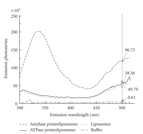
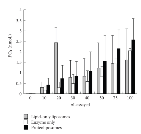
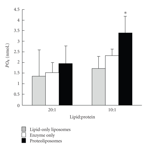
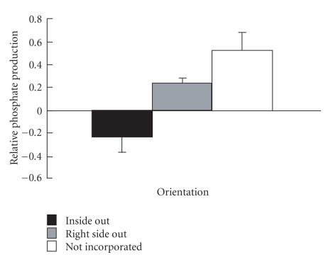
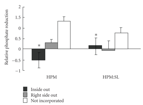
Sperm membranes change in structure and composition upon ejaculation to undergo capacitation, a molecular transformation which enables spermatozoa to undergo the acrosome reaction and be capable of fertilization. Changes to the membrane environment including lipid composition, specifically lipid microdomains, may be responsible for enabling capacitation. To study the effect of lipid environment on proteins, liposomes were created using lipids extracted from bull sperm membranes, with or without a protein (Na(+) K(+)-ATPase or α-amylase). Protein incorporation, function, and orientation were determined. Fluorescence resonance energy transfer (FRET) confirmed protein inclusion in the lipid bilayer, and protein function was confirmed using a colourometric assay of phosphate production from ATP cleavage. In the native lipid liposomes, ATPase was oriented with the β subunit facing the outer leaflet, while changing the lipid composition to 50% native lipids and 50% exogenous lipids significantly altered this orientation of Na(+) K(+)-ATPase within the membranes.


Figures

Figure 1
Figure 1
SEM micrograph of typical proteoliposomes. These proteoliposomes were made by high pressure nitrogen filtration, fixed on polished carbon planchettes with OsO4, dried, sputter coated with 15 nm argon, and viewed using a Hitachi S-570 Scanning Electron Microscope. The dotted scale bar across the bottom represents 860 nm. Arrow indicates typical liposome.
Figure 2
Figure 2
Fluorescence resonance energy transfer to detect ATPase incorporation. Typical fluorescence emission from a single scan of liposomes (dotted) and proteoliposomes containing amylase (dashed) or ATPase (solid) and buffer (dashed dotted). All liposomes were prepared by filtration, contained 2 moL% dansyl, and were excited at 278 nm, the wavelength of excitation for tryptophan endogenous in the enzymes, and emission recorded over 300–520 nm; each preparation was scanned 3 times within 20 minutes. Mean emission intensities obtained from the three scans at 500 nm (grey vertical line), the wavelength of maximum emission of dansyl when excited by emitted light from nearby tryptophan, differed significantly within each replicate (a, b, c, d; n = 3).
Figure 3
Figure 3
Mean phosphate (PO4) detected (nmoL ± SE) in liposomes (lipid-only liposomes), Na+ K+-ATPase alone (enzyme-only), and proteoliposomes (liposomes containing ATPase) made by filtration (n = 3). Measurements were taken at 0, 10, 20, 30, 40, 50, 75, and 100 μl. Liposomes and proteoliposomes contained 0.5 μg lipid/μl prior to vesicle production, while proteoliposomes and enzyme-only contained 0.025 μg Na+ K+-ATPase/μl. The PO4 in each well was detected by reading colour intensity at 750 nm. The slope across concentrations was linear with increasing concentration for enzyme-only and proteoliposomes (P < .001).
Figure 4
Figure 4
Mean phosphate (PO4) detected (nmoL ± SD) in liposomes (lipid-only liposomes), Na+ K+-ATPase alone (enzyme-only), and proteoliposomes (liposomes containing ATPase) made by filtration with a lipid : protein ratio of 20 : 1 (n = 4) or 10 : 1 (n = 4) after 60 minutes of incubation. Liposomes and proteoliposomes contained 0.5 μg lipid/μl prior to vesicle production, while proteoliposomes and enzyme-only contained 0.025 μg Na+ K+-ATPase/μl for 20 : 1 and 0.05 μg Na+ K+-ATPase/μl for 10 : 1. PO4 in each well was detected by reading colour intensity at 750 nm. Significant differences within lipid : protein are marked by ∗.
Figure 5
Figure 5
Orientation of Na+ K+-ATPase in proteoliposomes measured by the sidedness assay (n = 3). Proteoliposomes were created by nitrogen filtration, incubated ± ouabain ± detergent and then function (PO4 production from ATP) was measured and used to calculate sidedness [33] to determine the relative amount of Na+ K+-ATPase that was facing inside out, right side out, or was not incorporated. All three orientations of enzyme were statistically different from each other (P < .05).
Figure 6
Figure 6
Orientation of Na+ K+-ATPase in HPM alone and in HPM: SL proteoliposomes measured by the sidedness assay. Proteoliposomes were created by nitrogen filtration using HPM or HPM: SL lipids (1 : 1; wt : wt), incubated ± ouabain ± detergent and then function (PO4 production from ATP) was measured and used to calculate sidedness [33] to determine the relative amount of Na+ K+-ATPase that was facing inside out, right side out, or was not incorporated. Differences between HPM proteoliposomes and HPM: SL proteoliposomes are indicated by ∗.

Similar articles

Cited by

References

    1. Post H, Schwarz A, Brandenburger T, Aumüller G, Wilhelm B. Arrangement of PMCA4 in bovine sperm membrane fractions. International Journal of Andrology. 2010;33(6):775–783. - PubMed
    1. Edidin M. Lipids on the frontier: a century of cell-membrane bilayers. Nature Reviews Molecular Cell Biology. 2003;4(5):414–418. - PubMed
    1. Khalil MB, Chakrabandhu K, Xu H, et al. Sperm capacitation induces an increase in lipid rafts having zona pellucida binding ability and containing sulfogalactosylglycerolipid. Developmental Biology. 2006;290(1):220–235. - PubMed
    1. Yanagimachi R. Mammalian fertilization. In: Knobil E, Neill J, editors. The Physiology of Reproduction. Vol. 1. New York, NY, USA: Raven Press; 1994. pp. 189–317.
    1. Gadella BM, Tsai PS, Boerke A, Brewis IA. Sperm head membrane reorganisation during capacitation. International Journal of Developmental Biology. 2008;52(5-6):473–480. - PubMed

LinkOut - more resources