. 2011:2011:670479.
doi: 10.1155/2011/670479. Epub 2010 Sep 19.

Regulation of Serum Response Factor and Adiponectin by PPARγ Agonist Docosahexaenoic Acid

Affiliations

Regulation of Serum Response Factor and Adiponectin by PPARγ Agonist Docosahexaenoic Acid

Clayton Johnson et al. J Lipids. 2011.

Abstract

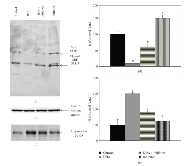
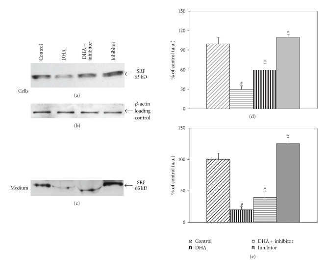
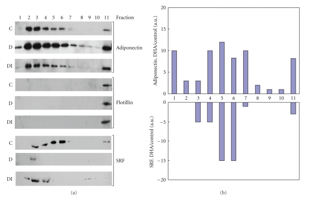
Recent studies indicate that significant health benefits involving the regulation of signaling proteins result from the consumption of omega-3 polyunsaturated fatty acids (ω-3 PUFAs). Serum response factor (SRF) is involved in transcriptional regulation of muscle growth and differentiation. SRF levels are increased in the aging heart muscle. It has not been examined whether SRF is made by adipocytes and whether SRF secretion by adipocytes is modulated by PPARγ agonist DHA. Adiponectin is made exclusively by adipocytes. We and others have previously reported that PUFAs such as DHA increase adiponectin levels and secretion in adipocytes. Here we show that DHA downregulates SRF with a simultaneous upregulation of adiponectin and that both these responses are reversible by PPARγ antagonist. Furthermore, there appears to be a direct relationship between DHA exposure and increased levels of membrane-associated high-density adiponectin, as well as lower levels of membrane-associated SRF. Thus, we find that the levels of SRF and adiponectin are inversely related in response to treatment with PPARγ agonist DHA. Decreased levels of SRF along with increase in membrane-associated adiponectin could in part mediate the health benefits of DHA.


Figures

Figure 1
Figure 1
Effects of DHA on SRF and adiponectin in 3T3-F442A adipocytes. 3T3-F442A adipocytes differentiated as described in Methods were treated with 25 μM DHA, 25 μM DHA plus 1.5 μM GW9662 or 1.5 μM GW9662 in DMEM containing 1% FBS for 16 h. Cells were lysed and separated on 10% SDS-PAGE followed by Western blotting with specific antibody. (a) Blots were analyzed with SRF C-terminus antibody. (d) Bar graph shows quantification using densitometric analysis of ≈65 kDa SRF protein. Data in the bar graph represents means of three independent experiments (*P < .025, # P < .05). (b) The same blot was analyzed with β-actin antibody to examine equal loading of proteins. (c) The same samples were also examined for the expression of adiponectin. Blots were probed with adiponectin antibody; bar graph shows quantitation of adiponectin protein using densitometric analysis and is expressed as percent of control. (e) Data in the bar graph represents means of three independent experiments (*P < .05, # P < .05).
Figure 2
Figure 2
SRF expression in mouse adipocytes treated with DHA. Equal volumes (1-2 ml) of freshly isolated adipocytes were incubated in DMEM-HEPES supplemented with penicillin-streptomycin, containing 1% BSA. Adipocytes were treated as specified control, 25 μM DHA, 25 μM DHA plus 1.5 μM GW9662, or 1.5 μM GW9662 alone for 16 h. Cells and medium were separated and analyzed for SRF expression. (a) Western blots of SRF expression in mouse adipocytes. (d) Data in the adjacent bar graph represents one of two experiments done in duplicate(*P < .03, # P < .05). (b) The blot from part A was probed with β-actin antibody. (c) Western blot of SRF expression in adipocyte conditioned medium. Densitometric analysis of SRF expression in cells, and medium is expressed as a percent of control. (e) The bar graph represents means of all replicate experiments (*P < .025, # P < .025).
Figure 3
Figure 3
Fractionation of adipocyte medium following treatment with DHA or DHA in the presence of PPARγ inhibitor GW 9662. Mouse adipocytes were treated: control (C), 25μM DHA (D), or 25 μM DHA plus 1.5 μM GW9662 (DI). Equal volumes of medium secreted from treatments were fractionated on sucrose gradients and separated into 11 equal fractions as described in Methods. (a) Equal volumes of each fraction were separated on SDS-PAGE gels and analyzed by Western blotting using adiponectin, Flotillin, or SRF antibody as described in Methods. (b) Bar graph represents densitometric analysis of change in adiponectin and SRF expression following DHA treatment in each fraction: DHA/Control.

References

    1. Haffner SM. Abdominal adiposity and cardiometabolic risk: do we have all the answers? American Journal of Medicine. 2007;120(9):S10–S16. - PubMed
    1. Posern G, Treisman R. Actin’ together: serum response factor, its cofactors and the link to signal transduction. Trends in Cell Biology. 2006;16(11):588–596. - PubMed
    1. Pipes GCT, Creemers EE, Olson EN. The myocardin family of transcriptional coactivators: versatile regulators of cell growth, migration, and myogenesis. Genes and Development. 2006;20(12):1545–1556. - PubMed
    1. Zhang X, Azhar G, Chai J, et al. Cardiomyopathy in transgenic mice with cardiac-specific overexpression of serum response factor. American Journal of Physiology—Heart and Circulatory Physiology. 2001;280(4):H1782–H1792. - PubMed
    1. Parlakian A, Charvet C, Escoubet B, et al. Temporally controlled onset of dilated cardiomyopathy through disruption of the srf gene in adult heart. Circulation. 2005;112(19):2930–2939. - PubMed

LinkOut - more resources