

Glomus tumor of the stomach simulating a gastrointestinal stromal tumor: a case report and review of literature
- PMID: 21490829
- PMCID: PMC3075157
- DOI: 10.1159/000112862
Glomus tumor of the stomach simulating a gastrointestinal stromal tumor: a case report and review of literature
Abstract
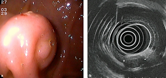
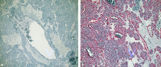
Glomus tumor is an infrequent and in most cases benign mesenchymal neoplasia which affects subcutaneous/submucosal tissue and occurs in the gastrointestinal tract, solid organs (e.g. liver, kidney) and the extremities. Visceral glomus tumor of the stomach generally presents with non-specific epigastric pain, loss of appetite and GI bleeding (melaena), often without haemodynamic instability. Macroscopic appearances on upper GI endoscopy are non-diagnostic. Endosonographic appearances are generally heterogenous and poorly-reflective, hence fail to differentiate glomus tumor from other potential diagnoses. Histological confirmation of the diagnosis is only possible when a fine needle biopsy is inclusive of abnormal tissue. These difficulties in diagnosis mean that in many cases, only immunohistochemical analysis of surgically resected tissue can distinguish glomus tumor from several possible differentials. Therefore, endoscopically-assisted laparoscopic curative wedge-resection of a lesion suspicious for glomus tumor of the upper gastrointestinal tract must be considered first-line in terms of a combined investigative and curative approach.
Keywords: GIST; Gastric glomangioblastoma; Glomus tumor.
Figures
References
-
- Caccamo D, Danedo M, Gordon RE. Glomus tumor of the stomach. Mt Sinai J Med. 1987;54:344–347. - PubMed
-
- De Busscher G. Les anatomoses arterioveineuses de l'estomac: An ultrastructural study. Acta Neurol Morphol. 1948;6:87–105. - PubMed
-
- Smol'iannikov A. Glomus tumors. Vopr Onkol. 1974;20:104–116. - PubMed
-
- Kirschbaum D, Teitelman L. Malignant tumor of the omentum simulating glomangioma. Arch Path. 1939;27:95.
-
- Yannopoulos K, Stout AP. Smoot muscle tumors in children. Cancer. 1962;15:958. - PubMed
Publication types
LinkOut - more resources
Full Text Sources
