. 2011 Apr 7;6(4):e18410.
doi: 10.1371/journal.pone.0018410.

Delayed re-epithelialization in periostin-deficient mice during cutaneous wound healing

Affiliations

Delayed re-epithelialization in periostin-deficient mice during cutaneous wound healing

Takashi Nishiyama et al. PLoS One. .

Abstract

Background: Matricellular proteins, including periostin, are important for tissue regeneration.

Methods and findings: Presently we investigated the function of periostin in cutaneous wound healing by using periostin-deficient ⁻/⁻ mice. Periostin mRNA was expressed in both the epidermis and hair follicles, and periostin protein was located at the basement membrane in the hair follicles together with fibronectin and laminin γ2. Periostin was associated with laminin γ2, and this association enhanced the proteolytic cleavage of the laminin γ2 long form to produce its short form. To address the role of periostin in wound healing, we employed a wound healing model using WT and periostin⁻/⁻ mice and the scratch wound assay in vitro. We found that the wound closure was delayed in the periostin⁻/⁻ mice coupled with a delay in re-epithelialization and with reduced proliferation of keratinocytes. Furthermore, keratinocyte proliferation was enhanced in periostin-overexpressing HaCaT cells along with up-regulation of phosphorylated NF-κB.

Conclusion: These results indicate that periostin was essential for keratinocyte proliferation for re-epithelialization during cutaneous wound healing.


Conflict of interest statement

Competing Interests: The authors have declared that no competing interests exist.

Figures

Figure 1
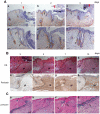
Figure 1. The expression of periostin in normal mouse and human skin.
(A) The mRNA expression of periostin by RNA in-situ hybridization (a, c) and the protein expression of periostin by immunohistochemistry (b, d) in normal mouse skin. Insets are high-magnification views of hair follicles. Periostin mRNA was expressed in the bulge region of the hair follicle, especially in the outer cells along the basement membrane (a, arrow) not in the inner cells (a, arrowhead), and its protein was localized at the basement membrane of the follicular bulge region (b, brown stain). In the epidermis, periostin mRNA was expressed in a patch-wise pattern in the cells along the basement membrane (c, arrow), and its protein was localized at the basement membrane (d, brown stain). Scale bars: 100 µm; epi: epidermis; hf: hair follicle; sg: sebaceous gland; der: dermis (B) The mRNA expression of periostin assessed by in situ hybridization (a, e); and the protein expression of periostin, by immunohistochemistry (b), together with H&E staining (c) and vimentin protein expression (d) in normal human skin. Figures c -f show a high magnification of the border area of the epidermis and the dermis. Immunohistochemistry was performed using anti-periostin antibodies (b, f) and anti-vimentin antibodies (d). Periostin mRNA was expressed only in the keratinocytes (arrows in “a” and “e”) on the basement membrane, but not in melanocytes (arrowhead in “d”) or fibroblasts (arrow in “d”), and periostin protein was localized in the basement membrane (b and f, brown stain). Scale bar: 50 µm; epi: epidermis; der: dermis.
Figure 2
Figure 2. Periostin expression during wound healing.
The quantitative RT-PCR for mouse periostin mRNA were performed. The wounds were removed at 1, 2, 3, 5, 7, 10 or 14 day after wounding. We used intact skin as a control tissue (con.). The representative result of RT-PCR bands is shown (A), and the quantitative result from 4 independent experiments is shown in a graph (B). Significant periostin expression was observed at day 3, and peaked at day 7. Bars represent the mean ± S.E.
Figure 3
Figure 3. Periostin protein expression in wound healing.
(A) Periostin protein expression in early wound healing. Wounds at day 1(a, d), 2(b, e), and 3(c, f) after wounding were stained with anti-periostin antibodies. Figures d, e, and f show a high magnification of “a,” “b,” and “c,” respectively. At day 1, periostin protein was expressed mainly in hair follicles (d, arrow) and weakly at the basement membrane in the hair follicle opening area (d, arrowhead). At days 2 and 3, the protein was localized more clearly at the basement membrane of the epidermis (b, c; arrow) and diffusely around the hair follicle (e, f; arrowhead). Red arrowheads indicate the wound edge. Scale bar: 100 µm (a, b), 50 µm (c, d); sc: scab. (B) H&E staining (a–d) and periostin protein expression in wounded skin assessed by immunohistochemistry (e–h) at 3, 5, 7, and 10 after wounding. Periostin was localized in granulation tissues. The dotted line in each photo indicates the border between the dermis and the granulation tissue. Scale bar: 100 µm; gt: granulation tissue (C) Periostin mRNA in situ hybridization of skin at 5 days after wounding. The sections were probed with the anti-sense cRNA probe (a) and the control sense probe (b). The periostin-positive area in “a” is magnified in “c and “d”: periostin-positive fibroblasts surrounding a hair follicle (arrowhead in “c”) and at the border of the wound area (arrow in “d”) are seen. The signals were not detected with the sense probe (b). Scale bar: 100 µm (a, b), 50 µm (c, d).
Figure 4
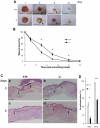
Figure 4. Delayed wound contraction in periostin−/− mice.
(A) Representative macroscopic views of skin wounds in the periostin WT (+/+, upper row) and −/− (lower row) mice on 1, 3, 4, and 8 days after wounding. This wound healing was monitored by taking digital photographs. Scale bar: 3 mm. Note the delay of wound healing in the −/− mice. The arrow in photos of days 3 and 5 of −/− mice shows a clearer border of the wound area compared with the border of that in the +/+ mice at 3 and 5 days (arrowhead). (B) The wound size is shown. Time-course of changes in wound closure ratio after wounding on 0, 1, 3, 5, 8 and 10 days. From the photographs, percent wound size was calculated by measuring the wounded area. A total of 20 wounds were assessed (Vertical lines represent the mean ± S.E. *P<0.005). (C) Delayed re-epithelialization in periostin/− mice. The wound area of WT (a, c) and periostin−/− (b, d) mice was excised on day 3 (a, b) or 5 (c, d) after wounding, sectioned, and stained with H&E. Arrowheads and arrows indicate the original wound edge and leading edge of the re-epithelialized area, respectively. The dotted area indicates the newly formed epidermis. Scale bar: 50 µm. (D) From the photographs, the percent re-epithelialization level of WT (a, c) and periostin−/− (b, d) mice was calculated by measurement of the re-epithelialized area at days 3 and 5 after wounding. Four wounds were assessed. Bars represent the mean ± S.E. *P<0.05, **P<0.01.
Figure 5
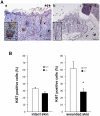
Figure 5. Reduced number of Ki67-positive cells in hair follicles in periostin-/- mice.
(A) These sections of a day-3 wound in WT (+/+) (a) and periostin/− mice (b) were stained with anti-Ki67 antibody. Insets show high-magnification views of a hair follicle. The cells in the granulation tissue of both +/+ and periostin/− mice were positively stained with anti-Ki67 antibody (arrowhead), but those in the hair follicle of periostin/− mice were weakly stained (inset). The dotted line indicates the border between the dermis and the granulation tissue. Scale bar: 50 µm, gt; granulation tissue (B) Ki67-positive cell index (percentage of positive cells among all keratinocytes) for the bulge region of hair follicles of intact skin or of wounded skin. A total of 6 hair follicles surrounding the wound were assessed. Bars represent the mean ± S.E. *P<0.05.
Figure 6
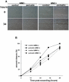
Figure 6. Periostin enhances re-epithelialization by inducing cell growth in vitro in the keratinocyte scratch assay.
(A) Cells of the human keratinocyte cell line HaCaT were transfected with the periostin-HA construct, and the resulting transfectants were tested by performing the keratinocyte scratch assay to determine the effect of periostin on the wound closure. This assay was performed in the presence or absence of MMC (mitomycin C) to detect the effect of cell proliferation on the wound closure. Representative wound-closing cells (34 hr) after onset of scratching (0 hr) are shown. (B) The level of wound closure was examined at 0, 12, 24, 34 and 48 hr, after scratching the cell sheet. Vertical lines represent the mean ± S.E.
Figure 7
Figure 7. Periostin induces cell proliferation via the NF-κB signaling.
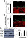
HaCaT transfectants of the periostin-HA expression vector or control vector were cultured to confluence, and the cultures were continued for 1 week in 4% FBS medium. Then, the cells were serum-starved for 24 hr. After that, the cells were stimulated with 10% FBS plus 50 nM BrdU for 12 hr (A) or with 10% FBS for 15 min (B). (A) The graph shows the mean percentage of BrdU-positive cells from 5 experiments. Representative photos of BrdU signals in control and periostin-HA transfectants are also shown (x400). The lower photos show the nuclear staining by PI (propidium iodide). Note that the number of BrdU-labeled cells was increased (about 2 fold) in the periostin-HA transfectants. Bars represent the mean ± S.E. *P<0.05. (B) The graph shows the mean number of phosphorylated NF-κB (pNF-κB: p-p65)-positive cells from 5 experiments, in which immunohistochemistry with anti-pNF-κB antibody was performed. Representative photos of pNF-κB signals in control and periostin-HA transfectants are shown (x400). PI-positive nuclei are seen in the lower photos. Note that the number of pNF-κB-positive cells was increased (about 2 fold) in the periostin-HA transfectant. Bars represent the mean ± S.E. *P<0.05. (C) NF-κB phosphorylation assessed by Western blotting of HaCaT transfectants. Cells were treated with FBS in the same manner as described in “B,” then sampled after 5, 10, and 30 min after FBS treatment, and blotted with anti-pNF-κB (phosphorylated p65), anti-NF-κB (non-phosphorylated p65), anti-phospho ERK (upper signal of pNF-κB), anti-ERK, and anti-β-actin antibodies. “C” indicates the control vector. Representative data from Western blot analysis from 3 independent experiments are shown. (D) The quantitative data for the ratio of pNF-κB (phosphorylated p65) to NF-κB (non-phosphorylated p65) obtained from 3 independent experiments are shown in this graph. Bars represent the mean ± S.E. In the periostin- over-expressing cells, NF-κB phosphorylation and the amount of ERK (arrows in “C”) were increased.
Figure 8
Figure 8. Enhancement of the proteolytic cleavage of laminin γ2 by its association with periostin.
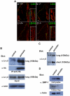
(A) Immunohistochemistry of laminin γ2 in hair follicles. Sections of WT (+/+) (a, c) and −/− hair follicles (b, d) were immunostained with 2 different anti-laminin γ2 antibodies, i.e., L4m, recognizing both the long and short forms of laminin γ2 (a, b), and LE4-6, which recognizes only the long form (c, d). The red color shows the nuclear staining by PI (propidium iodide). (B) Immunoprecipitation of periostin and laminin γ2. With anti-HA antibody periostin was immunoprecipitated from the periostin-HA HaCaT transfectants that expressed the long-form laminin γ2 (Input) and from the control cells treated with the vector only (control), and Western blot analysis was performed with anti-laminin γ2 antibody (1–97) on this immunoprecipitant. The long form of laminin γ2 was detected as well as the perostin signal. Anti-actin antibody detected actin as a control. (C) Western blot analysis of laminin γ2. The periostin-HA HaCaT transfectants highly produced the immature laminin γ2 (non-cleaved long form: 150 kDa) and the activated laminin γ2 (cleaved short form: 105 kDa), as detected by anti- laminin γ2 antibody (1–97). (D) BMP-1in cell lysates or culture supernatants (Culture Sup.) from the periostin-HA HaCaT transfectants and from the control cells treated with the vector only (control). The accumulation of BMP-1 in cell lysates from the periostin-HA HaCaT transfectants was increased. The proteins in total cell lysates and cell culture supernatants were each separated by SDS-PAGE, and blotted with anti-BMP-1; and the former were blotted with anti-β-actin antibodies.
Figure 9
Figure 9. Fibronectin localization in hair follicles of WT and periostin/− mice.
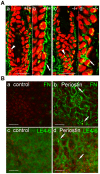
(A) Fibronectin expression (green fluorescence) was examined in hair follicles from WT (+/+; a, b) and periostin−/− mice (c, d) by using anti-fibronectin antibody. The red color shows the nuclear staining by PI. Photos “b” and “d” show high magnification views of “a” and “c,” respectively. Fibronectin was localized mainly at the basement membrane (b, arrow) and rarely in the cytoplasm in WT mice (a, arrowhead). However, in periostin−/− mice, the surface expression of fibronectin was rarely detected, and preferentially observed in the cytoplasm (c, arrowhead; d, arrow). Scale bar: 20 µm. (B) Fibronectin and laminin γ2 localization on the periostin HaCaT transfectant. The localization of fibronectin and laminin γ2 proteins on the cell surface of the periostin-HA (b, d) or the control vector (a, c). HaCaT transfectants were examined by using anti-fibronectin (a, b) and anti-laminin γ2 antibody (LE4-6; c, d). Fibronectin localization in periostin transfectants shows a patched pattern (b, arrow), consistent with that for laminin γ2 (LE4-6) in the same transfectants (d, arrow). Scale bar: 20 µm.

References

    1. Martin P. Wound healing-aiming for perfect skin regeneration. Science. 1997;276:75–87. - PubMed
    1. Eyden B. The myofibroblast, electron microscopy and cancer research. Int J Cancer. 2009;125:1743–1745. - PubMed
    1. Metcalfe AD, Ferguson MWJ. Tissue engineering of replacement skin: the crossroads of biomaterials, wound healing, embryonic development, stem cells and regeneration. J R Soc Interface. 2007;4:413–437. - PMC - PubMed
    1. Ito M, Liu Y, Yang Z, Nguyen J, Liang F, et al. Stem cells in the hair follicle bulge contribute to wound repair but not to homeostasis of the epidermis. Nature Med. 2005;11:1351–1354. - PubMed
    1. Howe A, Aplin AE, Alahari SK, Juliano RL. Integrin signaling and cell growth control. Curr Opin Cell Biol. 1998;10:220–231. - PubMed

Publication types

MeSH terms

LinkOut - more resources