. 2011 Apr 7;6(4):e18645.
doi: 10.1371/journal.pone.0018645.

The anti-metastatic nm23-1 gene is needed for the final step of mammary duct maturation of the mouse nipple

Affiliations

The anti-metastatic nm23-1 gene is needed for the final step of mammary duct maturation of the mouse nipple

Camille Deplagne et al. PLoS One. .

Abstract

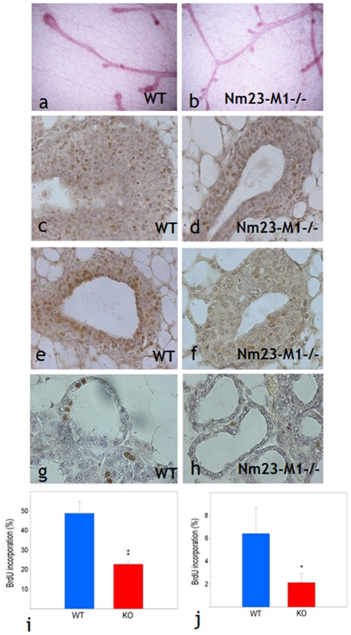
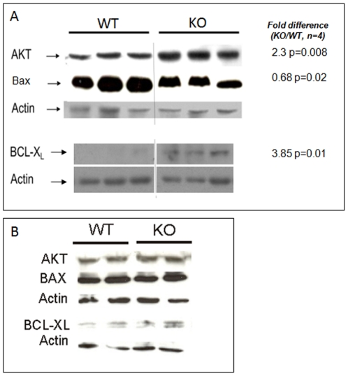
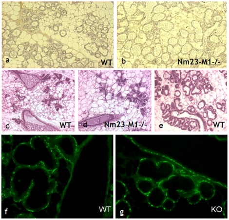
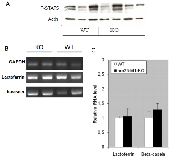
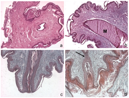
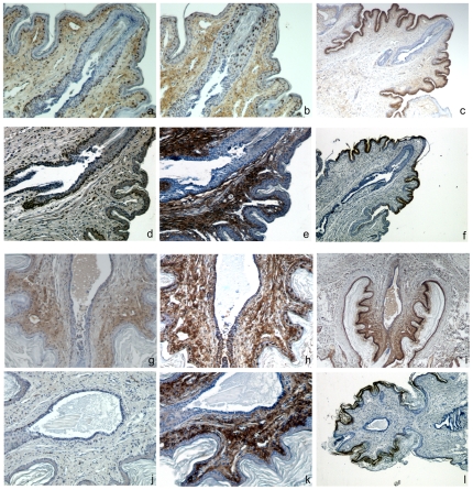
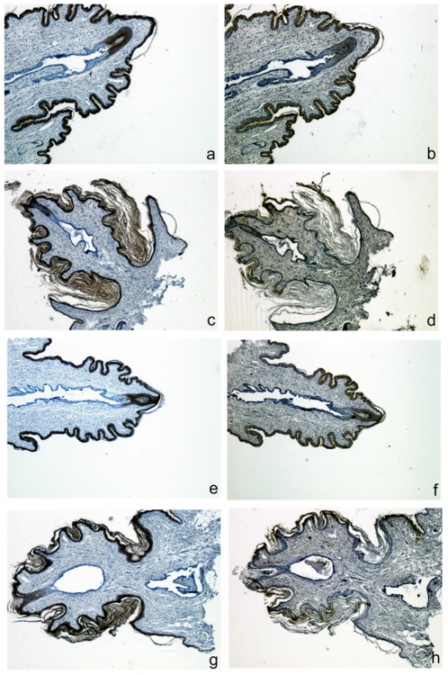
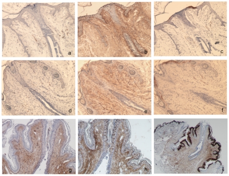
Nm23/NDP kinases are multifunctional enzymes involved in the general homeostasis of triphosphate nucleosides. Numerous studies have shown that NDPKs also serve as regulatory factors of various cell activities, not always connected to nucleotide phosphorylation. In particular, the nme-1 gene, encoding the NM23-1/NDPKA protein, has been reported as a metastasis suppressor gene. This activity was validated in hepatocellular tumors induced in nm23-1 deficient mice. Yet, data describing the primary physiological functions of nm23-1/NDPKA is still scarce. We have characterized in depth the phenotype of nm23-1 deletion in the mammary gland in mice carrying whole body nm23-M1 invalidation. We also asked why the nm23-M1⁻/⁻ mutant females displayed severe nursing disability. We found that the growth retardation of mutant virgin glands was due to reduced proliferation and apoptosis of the epithelial cells within the terminal end buds. The balance of pro/anti-apoptotic factors was impaired in comparison with wild type glands. In the lactating glands, the reduced proliferation rate persisted, but the apoptotic factors were unchanged. However, those defects did not seem to affect the gland maturation since the glands lacking nm23-1/NDPKA appeared morphologically normal. Thorough examination of all the functional aspects of the mammary glands revealed that lack of nm23-1/NDPKA does not impact the production or the ejection of milk in the lumen of lobuloalveolae. Interestingly, an epithelial plug was found to obstruct the extremity of the unique lactiferous duct delivering the milk out of the nipple. These cells, normally disappearing after lactation takes place, persisted in the mutant nipples. This work provides a rare instance of nm23-1/NDPKA physiological functions in the mammary glands and reveals its implication as a modulator factor of proliferation and apoptosis in this tissue.


Conflict of interest statement

Competing Interests: The authors have declared that no competing interests exist.

Figures

Figure 1
Figure 1. Lack of nm23-M1 results in impaired proliferation.
Whole mounts of WT (a) and nm23-M1−/− (b) mammary glands show a delayed growth of the TEBs in the mutant glands (original magnification X5). BrdU incorporation (brown nuclei) was evidenced in sections from mammary glands from 6 weeks old WT (c,e) or nm23-M1−/− (d,f) virgin females and L1 WT (g) or L1 nm23-M1-/- (h) lactating females (original magnification X400). Quantifications in virgin glands (i) and lactating glands (j) revealed a drop in BrdU incorporation in the epithelial cells of the mutant glands. KO: nm23-M1−/−.
Figure 2
Figure 2. Lack of nm23-M1 results in diminished apoptosis markers in the virgin but not the lactating mutant glands.
A: protein extracts from 6 weeks old virgin WT and nm23-M1−/− (KO) glands were processed for western-blotting. Images were analyzed by densitometry to measure intensity differences between the two groups. Results are indicated on the right hand side. B: protein extracts from L1 lactating WT and nm23-M1−/− (KO) glands were processed for western-blotting. No difference in band intensity was evidenced between the two groups.
Figure 3
Figure 3. The morphology of lactating L1 and L3 glands seems normal in mutant females.
Sections from lactating L1 WT (a) and nm23-M1−/− (b) mammary glands do not reveal any abnormality. L3 nm23-M1−/− glands (d) show signs of involution as evidenced by the diminished density of lobuloalveolar structures, not seen in L3 WT lactating gland (e). However, L3 glands from WT females from which the litters have been removed (c) do not appear different from nm23-M1−/− L3 gland (Original magnification X200). Sections have been stained with anti-alpha actin to reveal normal pattern of myoepithelial cells in WT (f) and mutant (g) L1 lactating glands (Original magnification X400).
Figure 4
Figure 4. Mutant mammary glands do not present functional defects.
Western-blots have been carried out to detect the level of phospho-STAT5. Actin was detected to check on sample loading. (A) RT-PCRs have been performed and quantified by densitometry. RNA levels related to GAPDH signal are reported (C).
Figure 5
Figure 5. Nipples from nm23-M1−/− mammary glands are obstructed.
Sections from WT (a, c) and nm23-M1−/− (b, d) nipples were sectioned and stained by hematoxylin and eosin (a, b) or trichrom Masson (c, d). The general architecture of dermis and epidermis of the mutant nipple doesn't seem overly changed. However, close examination revealed that the final opening of the lactiferous canal is obstructed by epithelial cells in the mutant glands (arrow). M: milk. Original magnification X200 (a, b) X400 (c,d).
Figure 6
Figure 6. Proliferation and elimination of the epithelial plug does not occur in the lactiferous sinus of nipples from females which are not nursing.
Serial sections of nipples were stained with anti-PCNA (a, d, g and j), anti-beta-galactosidase (b, e, h and k) and anti-K2e (c, f, i and l). Sections from mutant (a, b and c) and WT (d, e and f) nipples from nursing females are fully opened at the edge and show numerous PCNA-positive cells within the wall of the lactiferous sinus. Sections from mutant (g, h and i) and WT (j, k and l) nipples from non nursing females show a persisting epithelial plug with numerous beta-galactosidase-positive cells (h), accumulation of milk in the lactiferous sinus and only a few PCNA-positive cells. Note the intense beta-galactosidase staining in the keratinocytes. Original magnification X75 (a, b, d, e, g, h, j and k) X150 (c, f, I and l).
Figure 7
Figure 7. Nipples from nm23-M1-/- females present normal epidermis.
Serial sections of nipples were stained with anti-K10 (a, c, e and g) and anti-K14 (b, d, f and h). Sections from WT (a, b, c and d) and mutant (e, f, g and h) nipples show no difference in K10 or K14 signal, whether they were retrieved from nursing (a, b, e and f) or not nursing (c, d, g and h) females. Original magnification X75.
Figure 8
Figure 8. Nipples from nm23-M1+/− virgin females present an epithelial plug rich in beta-galactosidase positive cells.
Serial sections of nipples from 6 weeks old (a, b and c), 12 weeks old (d, e and f) and L1 lactating (g, h and i) nm23-M1+/− females were stained with anti-PCNA (a, d, and g), anti-beta-galactosidase (b, e and h) and anti-K2e (c, f and i). Sections from 6 and 12 weeks old display an epithelial plug at the edge of the lactiferous sinus with numerous apoptotic figures (a) and beta-galactosidase-positive cells (b,e). Very few PCNA-positive cells could be seen (a, d). In the nipple from L1 lactating heterozygous females, PCNA-positive cells appeared more numerous and beta-galactosidase-positive cells disappeared together with the epithelial plug. Original magnification X150 except for i: X75.

Similar articles

Cited by

References

    1. Boissan M, Dabernat S, Peuchant E, Schlattner U, Lascu I, et al. The mammalian Nm23/NDPK family: from metastasis control to cilia movement. Mol Cell Biochem. 2009;329:51–62. - PubMed
    1. Steeg PS, Bevilacqua G, Sobel ME, Liotta LA. Identification and characterization of differentially expressed genes in tumor metastasis: the nm23 gene. Basic Life Sci. 1991;57:355–360; discussion 360-351. - PubMed
    1. Steeg PS, Bevilacqua G, Kopper L, Thorgeirsson UP, Talmadge JE, et al. Evidence for a novel gene associated with low tumor metastatic potential. J Natl Cancer Inst. 1988;80:200–204. - PubMed
    1. Steeg PS, Horak CE, Miller KD. Clinical-translational approaches to the Nm23-H1 metastasis suppressor. Clin Cancer Res. 2008;14:5006–5012. - PMC - PubMed
    1. Boissan M, Wendum D, Arnaud-Dabernat S, Munier A, Debray M, et al. Increased lung metastasis in transgenic NM23-Null/SV40 mice with hepatocellular carcinoma. J Natl Cancer Inst. 2005;97:836–845. - PubMed

Publication types

Substances