Review
. 2011:7:143-51.
doi: 10.2147/VHRM.S16328. Epub 2011 Mar 9.

Skeletonized coronary arteries: pathophysiological and clinical aspects of vascular calcification

Affiliations
Review

Skeletonized coronary arteries: pathophysiological and clinical aspects of vascular calcification

Carlos V Serrano Jr et al. Vasc Health Risk Manag. 2011.

Abstract

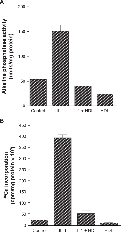
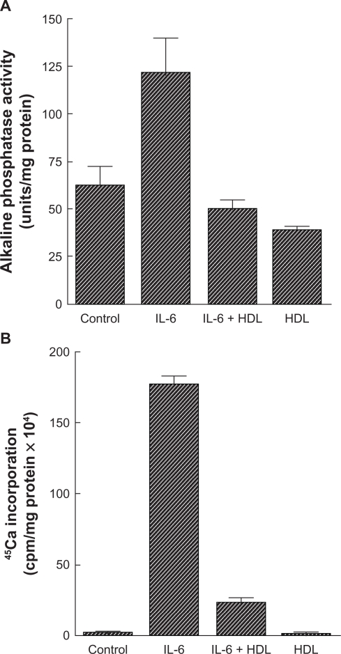
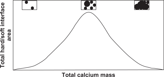
The role of calcification in coronary artery disease is gaining importance, both in research studies and in clinical application. Calcified plaque has long been considered to be the most important atherosclerotic plaque within the arterial tree and frequently presents a challenge for percutaneous intervention. Current investigations have shown that plaque calcification has a dynamic course that is closely related to the magnitude of vascular inflammation. Numerous inflammatory factors synthesized during the early stages of atherosclerosis induce the expression and activation of osteoblast-like cells localized in the arterial wall that produce calcium. There is no doubt that the role of these factors in calcification associated with coronary artery disease could be a crucial strategic point in prevention and treatment. A number of diagnostic imaging methods have been developed in recent years, but their performance needs to be improved. In this context, we undertook an update on coronary calcification, focusing on physiopathology, clinical implications, and imaging techniques.

Keywords: atherosclerotic plaques; vascular calcification; vascular smooth muscle cells.


Figures

Figure 1
Figure 1
Effect of high-density lipoprotein on cardiovascular cell responses to interleukin-1. A) Cardiovascular cell cultures were pretreated for 24 hours with high-density lipoprotein (200 μg/mL) in Dulbecco’s Modified Eagle Medium containing 5% fetal bovine serum, followed by the addition of fresh high-density lipoprotein with or without interleukin-1 (10 ng/mL). After two days, alkaline phosphatase activity was measured as previously described. Results from a representative of three experiments are shown as mean ± standard deviation of quadruple determinations. P < 0.005 for untreated control versus interleukin-1-treated samples, for interleukin-1- versus interleukin-1 + high-density lipoprotein-treated samples, and for untreated control versus high-density lipoprotein-treated samples. B) Cells were treated as described above. After 10 days, a 45Ca incorporation assay was performed. Results from a representative of three experiments are shown as mean ± standard deviation of quadruple determinations. P < 0.005 for untreated control versus interleukin-1-treated samples and for interleukin-1- versus interleukin-1 + high-density lipoprotein-treated samples. Adapted from Parhami et al.
Figure 2
Figure 2
Effect of high-density lipoprotein on cardiovascular cell responses to interleukin-6. A) Cardiovascular cell cultures were pretreated for 24 hours with high-density lipoprotein (200 μg/mL) in Dulbecco’s Modified Eagle Medium containing 5% fetal bovine serum, followed by the addition of fresh high-density lipoprotein with or without interleukin-6 (50 ng/mL). After four days, alkaline phosphatase activity was measured as previously described. Results from a representative of three experiments are shown as mean ± standard deviation of quadruple determinations. P < 0.001 for control versus interleukin-6-treated samples and for interleukin-6- versus interleukin-6 + high-density lipoprotein-treated samples; P < 0.05 for control versus high-density lipoprotein-treated samples. B) Cells were treated as described above. After 10 days, a 45Ca incorporation assay was performed. Results from three representative experiments are shown as mean ± standard deviation of quadruple determinations. P < 0.001 for control versus interleukin-6-treated samples and for interleukin-6- versus interleukin-6 + high-density lipoprotein-treated samples. Adapted from Parhami et al.
Figure 3
Figure 3
Relationship between plaque interface area versus total calcium mass. Biomechanical principles suggest the risk of plaque rupture should correlate with the interface area, which eventually decreases as calcified plaques begin to coalesce. The corresponding figure insets illustrate how the interface (the circumference around the black area) eventually decreases as calcified areas continue to form and grow. Adapted from Abedin et al.
Figure 4
Figure 4
Echocardiography image showing calcified aortic valve.
Figure 5
Figure 5
Intravascular ultrasound image showing necrotic core in a thin-cap fibroatheroma lesion appearing as red (top) and dense calcium in fibrocalcific disease appearing as white (bottom). Adapted from Murray et al.
Figure 6
Figure 6
Computed tomographic image showing coronary calcification not shown in the same lesion on coronary angiography. Adapted from Mittal et al.

Similar articles

Cited by

References

    1. Frink RJ, Achor RW, Brown AL, Jr, Kincaid OW, Brandenburg RO. Significance of calcification of the coronary arteries. Am J Cardiol. 1970;26:241–247. - PubMed
    1. Moses JW, Carlier S, Moussa I. Lesion preparation prior to stenting. Rev Cardiovasc Med. 2004;5(Suppl 2):S16–S21. - PubMed
    1. Abedin M, Tintut Y, Demer LL. Vascular calcification: Mechanisms and clinical ramifications. Arterioscler Thromb Vasc Biol. 2004;24:1161–1170. - PubMed
    1. Dhore CR, Cleutjens JP, Lutgens E, et al. Differential expression of bone matrix regulatory proteins in human atherosclerotic plaques. Arterioscler Thromb Vasc Biol. 2001;21:1998–2003. - PubMed
    1. Shanahan CM, Cary NR, Metcalfe JC, Weissberg PL. High expression of genes for calcification-regulating proteins in human atherosclerotic plaques. J Clin Invest. 1994;93:2393–2402. - PMC - PubMed