. 2011 Apr;7(4):e1001361.
doi: 10.1371/journal.pgen.1001361. Epub 2011 Apr 7.

The exocyst protein Sec10 interacts with Polycystin-2 and knockdown causes PKD-phenotypes

Affiliations

The exocyst protein Sec10 interacts with Polycystin-2 and knockdown causes PKD-phenotypes

Ben Fogelgren et al. PLoS Genet. 2011 Apr.

Abstract

Autosomal dominant polycystic kidney disease (ADPKD) is characterized by formation of renal cysts that destroy the kidney. Mutations in PKD1 and PKD2, encoding polycystins-1 and -2, cause ADPKD. Polycystins are thought to function in primary cilia, but it is not well understood how these and other proteins are targeted to cilia. Here, we provide the first genetic and biochemical link between polycystins and the exocyst, a highly-conserved eight-protein membrane trafficking complex. We show that knockdown of exocyst component Sec10 yields cellular phenotypes associated with ADPKD, including loss of flow-generated calcium increases, hyperproliferation, and abnormal activation of MAPK. Sec10 knockdown in zebrafish phenocopies many aspects of polycystin-2 knockdown-including curly tail up, left-right patterning defects, glomerular expansion, and MAPK activation-suggesting that the exocyst is required for pkd2 function in vivo. We observe a synergistic genetic interaction between zebrafish sec10 and pkd2 for many of these cilia-related phenotypes. Importantly, we demonstrate a biochemical interaction between Sec10 and the ciliary proteins polycystin-2, IFT88, and IFT20 and co-localization of the exocyst and polycystin-2 at the primary cilium. Our work supports a model in which the exocyst is required for the ciliary localization of polycystin-2, thus allowing for polycystin-2 function in cellular processes.


Conflict of interest statement

The authors have declared that no competing interests exist.

Figures
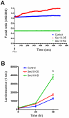
Figure 1
Figure 1. Sec10 knockdown in MDCK cells leads to ADPKD-like phenotypes.
A) Intracellular calcium levels were plotted as the ratio of Fura-2 fluorescence 340/380 nm, as a function of time in seconds. Confluent cells were exposed to a constant shear fluid flow rate of 5 ml/minute beginning at time = 0 seconds (arrow). Intracellular calcium and response to flow were decreased in Sec10 knockdown cells (green) and increased in Sec10-overexpressing cells (red), compared to wild-type MDCK cells (blue). B) Cell proliferation at 24 and 48 hours was plotted based on a cell titer luminescence assay, as a function of time, and was increased in Sec10 knockdown cells (* = p<0.01).
Figure 2
Figure 2. Sec10 knockdown in MDCK cells leads to activation of MAPK.
(A) Increased active, or phosphorylated, ERK (pERK) levels are seen by Western blot following Sec10 knockdown (Sec10-KD) in MDCK cells. (B) Quantification of results in (A) (** p<0.001). (C) Increased pERK in Sec10-KD cells is completely blocked by addition of the MEK inhibitor U0126 and the src inhibitor PP2, while the cAMP-activated PKA inhibitor H-89 reduces pERK levels to approximately normal levels. Other kinase inhibitors, including the PKC inhibitor bisindolylmaleimide I (BIM), and the mTOR inhibitor Rapamycin (RAP), showed no effect on pERK levels in these cells. (D) Quantification of results in (C) (* p<0.01, ** p<0.001). Experiments were run in triplicate, and equal amounts of protein, as determined by BCA, were loaded in each lane. Antibodies against pERK and total ERK (ERK) were used. The densities of the pERK bands were normalized to the densities of the corresponding total ERK bands, and the ratio of pERK:ERK was normalized to control MDCK cells.
Figure 3
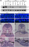
Figure 3. sec10MO embryos show abnormal pronephric development.
(A) Immunoblots showing the 1-3 dpf time course of zfSec10 knockdown in sec10MO embryos. sec10MO injections can lead to abnormal-appearing embryos (Table 1), but in this blot, 3 dpf sec10MO embryo lysates were isolated only from embryos without any obvious morphological defects. This demonstrates that these embryos still have strong knockdown of the zfSec10 protein. Lysates from abnormal-appearing embryos showed similar levels of zfSec10 knockdown (data not shown). 5 embryos loaded per lane. Positive control for Sec10 was from human Sec10 (hSec10) overexpressing MDCK cell lysates. Blot was probed with antibodies against hSec10, and gamma-tubulin as a loading control. (B-C′) Immunofluorescence with antibody against acetylated-tubulin (green), and the nuclear Hoechst stain (blue). Flattened Z-series from confocal imaging of medial (B,C) and posterior kidney (B′,C′), 24 hours post fertilization (hpf), lateral view, 80x magnification. Pronephric cilia length is similar between uninjected embryos (B/B′) and 15ng sec10MO embryos (C/C′); however, cilia within the medial pronephros are disordered (compare B and C). (D-E′) JB-4 resin section (with enlarged inset) of glomerular region, stained with Hematoxylin and Eosin, 3 dpf, transverse 4 μm section, 40x magnification. Wild-type embryos show an organized U-shaped glomerulus (D/D′), while 15ng sec10MO embryos show disorganization (E/E′).
Figure 4
Figure 4. Knockdown of sec10 partially phenocopies loss of pkd2.
(A-F) Gross phenotypes of zebrafish embryos at 3 dpf, lateral view, 4x magnification. Uninjected embryo (A), 4ng pkd2MO embryo with a severe curly tail up (B), 15ng sec10MO embryo with a moderate curly tail up (C). A synergistic interaction resulting in severe curly tail up was observed upon co-injection of sub-optimal doses of 0.25/2ng pkd2 MO +7.5ng sec10MO (D)—which do not result in curly tail up when injected alone (E, F). (G-I′) in situ hybridization for wt1a (with enlarged insets), 3 dpf, dorsal view, 16x magnification. An uninjected embryo with condensed glomerular stain (G/G′), a 4ng pkd2MO embryo with severe enlargement (H/H′), and a 15ng sec10MO embryo with severe enlargement (I/I′). (J) Increased phospho-ERK levels detected by Western blot in 4ng pkd2MO and 8+8ng sec10MO embryos at 5 dpf. One blot, loaded at 2 embryos per lane, was probed with antibody against phospho-ERK, then with antibody against total-ERK. The other blot, with the same lysates as above loaded at 10 embryos per lane, was probed with antibody against hSec10, then with antibody against gamma-tubulin.
Figure 5
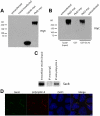
Figure 5. Sec10 biochemically interacts with polycystin-2.
(A) Human PKD2-myc in pcDNA3 was transfected using lipofectamine into HEK293 cells. Western blot was performed on the lysate (labeled “Pkd2-myc”), using antibody against the myc epitope tag. Polycystin-2-myc is seen, but at a higher molecular weight than expected, suggesting that the polycystin-2-myc is in a polymeric form. Identical results were seen using a monoclonal antibody against polycystin-2 (data not shown). “Cdc42-myc” = lysate from MDCK cells expressing Cdc42-myc (a positive control for the Western blot). (B) Purified Sec10-GST, but not GST alone, pulled down polycystin-2-myc from PKD2-myc transfected HEK293 cell lysate. (C) Exocyst Sec8 co-immunoprecipitated with polycystin-2, but not the isotype control, from intracellular vesicles isolated from mouse kidney lysate. The lanes in (C) were all from the same gel, though the intracellular vesicle input lane was separated from the other lanes (denoted by a white line). (D) Immunofluorescence staining, using a monoclonal antibody against exocyst Sec8 (green) and a polyclonal antibody against polycystin-2 (red), demonstrated co-localization of endogenous exocyst and polycystin-2 at the primary cilium (yellow in the merged panel) in MDCK cells grown on a Transwell filter for ten days. The panel showing DAPI-stained cell nuclei (blue) was taken at a different level inside the cell than the panels for Sec8 and polycystin-2, and is included here to delineate individual cells. Bar = 1 μm.
Figure 6
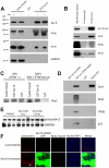
Figure 6. Sec10 interacts with IFT88 and IFT20.
(A) After incubation with HEK293 cell lysate, Sec10-GST, but not GST alone, pulled down the cilia transport proteins IFT88 and IFT20. As a positive control for exocyst binding, Sec8 is shown to bind to Sec10-GST. To demonstrate specificity for the pulldown products, we blotted for GAPDH, a house-keeping protein not known to interact with the exocyst. GAPDH was identified only in cell lysate, and not in pulldown fractions. (B) IFT88 and IFT20 co-immunoprecipitated with Sec10, but not isotype controls, from lysates of ciliated MDCK cells expressing a myc epitope-tagged Sec10. (C) To determine if IFT88 was necessary for the interaction between polycystin-2 and Sec10, Sec10-GST pulldowns were performed using lysate from both IFT88-deficient (94D), and IFT88-replete (BAP2) cell lines. There was no difference in the amount of polycystin-2 pulled down from the IFT-deficient or -replete cell lines. (D) In vitro translation of Sec8, polycystin-2, IFT88, and p53, followed by Sec10-GST pulldown, was also performed and an interaction was only detected between Sec8 and Sec10. This suggests that the Sec10/polycystin-2 and Sec10/IFT88 interactions are indirect. (E) Equal amounts of polycystin-2 protein, as determined by Western blot, are seen in control (T23), Sec10-overexpressing (A1), and Sec10 knockdown (10-4) MDCK cells. GAPDH staining is presented as a loading control. (F) Immunofluorescence staining, using a monoclonal antibody against acetylated alpha tubulin, that is specific for primary cilia (purple), and a polyclonal antibody against polycystin-2 (red), demonstrated co-localization of polycystin-2 at the primary cilia in control cells. As we previously reported , no, or few, primary cilia were seen in Sec10 knockdown cells, and the polycystin-2 appeared to be widely dispersed inside these cells. We occasionally saw concentrated polycystin-2 at what appeared to be the basal body or primary cilium (arrow); however, this occurred in cells demonstrating lower or no GFP expression, indicating a lack of Sec10 knockdown in those cells. The images presented are all X-Y sections, and were obtained using the same confocal settings. Please note that the DAPI staining for the cell nuclei was captured at a different level in the cell than the other sections and is included in the merge to delineate cell boundaries. Bar = 5 μm.
Figure 7
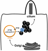
Figure 7. Model for the role of the exocyst in trafficking ciliary proteins.
Our model for the trafficking of essential proteins to the primary cilium posits that the exocyst is first localized to the primary cilium, and then targets and docks secretory vesicles from the trans-Golgi network carrying ciliary proteins such as polycystin-2 and IFT88—which are marked by the presence of IFT20.

Similar articles

Cited by

References

    1. Smyth BJ, Snyder R, Balkovetz DF, Lipschutz JH. Recent advances in the cell biology of polycystic kidney disease. In: Jeon KW, editor. Int Rev Cytol. San Diego: Elsevier Inc; 2003. pp. 52–89. - PubMed
    1. Consortium TIPD. Polycystic kidney disease: The complete structure of the PKD1 gene and its protein. Cell. 1995;81:289–298. - PubMed
    1. Mochizuki T, Wu G, Hayashi T, Xenophontos SL, Veldhuisen B, et al. PKD2, a gene for polycystic kidney disease that encodes an integral membrane protein. Science. 1996;272:1339–1342. - PubMed
    1. Gonzalez-Perrett S, Kim K, Ibarra C, Damiano AE, Zotta E, et al. Polycystin-2, the protein mutated in autosomal dominant polycystic kidney disease (ADPKD), is a Ca2+-permeable nonselective cation channel. Proc Natl Acad Sci U S A. 2001;98:1182–1187. - PMC - PubMed
    1. Vassilev PM, Guo L, Chen XZ, Segal Y, Peng JB, et al. Polycystin-2 is a novel cation channel implicated in defective intracellular Ca(2+) homeostasis in polycystic kidney disease. Biochem Biophys Res Commun. 2001;282:341–350. - PubMed

Publication types

MeSH terms