. 2011 Apr;7(4):e1002030.
doi: 10.1371/journal.pgen.1002030. Epub 2011 Apr 7.

Survival motor neuron protein regulates stem cell division, proliferation, and differentiation in Drosophila

Affiliations

Survival motor neuron protein regulates stem cell division, proliferation, and differentiation in Drosophila

Stuart J Grice et al. PLoS Genet. 2011 Apr.

Abstract

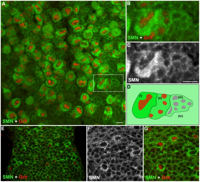
Spinal muscular atrophy is a severe neurogenic disease that is caused by mutations in the human survival motor neuron 1 (SMN1) gene. SMN protein is required for the assembly of small nuclear ribonucleoproteins and a dramatic reduction of the protein leads to cell death. It is currently unknown how the reduction of this ubiquitously essential protein can lead to tissue-specific abnormalities. In addition, it is still not known whether the disease is caused by developmental or degenerative defects. Using the Drosophila system, we show that SMN is enriched in postembryonic neuroblasts and forms a concentration gradient in the differentiating progeny. In addition to the developing Drosophila larval CNS, Drosophila larval and adult testes have a striking SMN gradient. When SMN is reduced in postembryonic neuroblasts using MARCM clonal analysis, cell proliferation and clone formation defects occur. These SMN mutant neuroblasts fail to correctly localise Miranda and have reduced levels of snRNAs. When SMN is removed, germline stem cells are lost more frequently. We also show that changes in SMN levels can disrupt the correct timing of cell differentiation. We conclude that highly regulated SMN levels are essential to drive timely cell proliferation and cell differentiation.


Conflict of interest statement

The authors have declared that no competing interests exist.

Figures

Figure 1
Figure 1. Growth defects in the SMN mutant CNS.
Prospero staining in a wild-type (A) and smnB (B) mutant CNS at the same scale. Both CNS are taken from larvae with 3rd instar spiracles and mouth hooks (wild-type, 4.5 Days old; smnB 5.5 days old). The smnB CNS is considerably reduced in size and is comparable to that of a 2nd instar wild-type. Both the thoracic ganglion and the brain lobes have retarded growth. Scale bars, 50 µm.
Figure 2
Figure 2. SMN is enriched in the postembryonic neuroblast (pNB) and forms a gradient in the developing neurons.
(A) A section of the 3rd instar thoracic ganglion showing SMN enriched in pNBs labelled with the pNB specific transcription factor Grh. The hatched box shows the pNB and daughter cells in (B) and (C) A gradient of SMN from pNBs through to the daughter cells in the same clone. (D) A schematic of the SMN gradient from (i) the most intense to (iv) the least intense. (E–G) SMN is enriched in pNBs of a late 1st instar larvae. (F, G) A separate scan of the pNBs in E. The antibody used was rabbit anti-SMN (gift from Jianhua Zhou 1∶2000). Scale bars, 10 µm.
Figure 3
Figure 3. Miranda localisation is abnormal in smn mutant neuroblasts.
Wild-type (A, B), smnA (C, D), and smnB (E, F) post embryonic neuroblasts (pNBs) showing Miranda and PH3 staining. During metaphase Miranda forms a crescent along the membrane of the neuroblast (A, B). In the smn mutant Miranda staining is reduced at the metaphase membrane (C and F) or can appear punctuate in the cytoplasm of the pNB (D). (G) Quantification of Miranda mislocalisation for each genotype. pNBs with inaccurate localisation, low level or the punctate localisation of Miranda were scored. Scale bar, 10 µm.
Figure 4
Figure 4. MARCM analysis of SMN in the larval CNS.
(A, B) Single frame images of a smnA MARCM clone showing the reduction of SMN. (C) Quantitative analysis of cell numbers in MARCM clones. Cell number counts at 65 hrs (wild-type, µ = 18; smnA, µ = 17), 82 hrs (wild-type, µ = 38.3; smnA, µ = 21.8; P<0.005, T-Test, n = 86) and 96 hrs (wild-type µ = 56.1; smnA, µ = 34.2; P<0.001, T-Test, n = 95). smnA clones show a significant reduction of cell proliferation over time. (D, E) Confocal pictures of a 64-hr wild-type and smnA clone. smnA clones have an abnormal shape, with GMCs formed in a inconsistent pattern. Clones imaged at 64 hrs. (F, G) Wild-type and smnA mutant MARCM clones stained for PH3. (H) The number of wild-type and smnA clones with pNBs positive for pH3 were counted and displayed as a percentage of the total number of clones from each type (wild-type  = 48%, n = 54; smnA  = 24% n = 56; P<0.001, T-Test). (I, J) smnA clones were stained for Grainyhead (Grh) to detect the presence of a neuroblast. (I) The majority of smnA clones have a large Grh positive cell present. Scale bars, 10 µm.
Figure 5
Figure 5. snRNP levels are reduced in SMN mutant MARCM clones.
(A–H) U2 and U5 expression in wild-type and smnA pNB MARCM clones using in situ hybridisation. Each clone is only one segment of a z-stack. Clones are labelled with GFP. U2 and U5 expression is located in the nucleus of wild-type clones (U2: A,B; U5: E, F) and in non mutant cells. SMN levels are reduced in smnA mutant clones (U2: C, D; U5: G, H). Scale bar, 10 µm.
Figure 6
Figure 6. An SMN gradient in the Drosophila adult testis.
(A) Schematic of the larval testis showing the position of germline stem cells (GSC), somatic stem cells (SSC), gonialblast (GB) and spermatogonia. (B–D) In Drosophila testis SMN staining is localised to the apical tip. (E–G) SMN is enriched in GSCs and levels decrease as cells differentiate. (H) Schematic showing the gradient in the testis from (i) the most intense to (v) the least intense. HTS (hu-li tai shao) labels the spectrosomes. The antibody used was rabbit anti-SMN (gift from Jianhua Zhou 1∶2000). Scale bars, 10 µm.
Figure 7
Figure 7. SMN is essential for the maintenance of male germline stem cells.
(A, B) smnA mitotic clone in the Drosophila adult testis. Testes with GFP negative goniablasts and spermatocytes (A, *) but no GFP negative stem cell are scored. Stem cells are recognised by their proximity to the hub (B, *, FasIII positive). (C) Stem cells are lost at a greater rate in smnA mitotic clone testis when compared to a wild-type control. Stem cells were counted in control and smnA testis at 3 (control, n = 55; smnA, n = 56) and 11 (control, n = 72; smnA, n = 78) days. There was very significant stem cell loss in smnA at 11 days (p<0.001, T-test) when compared to the control. (D–G) U2 levels are reduced in smnA mitotic clones. The hatched region shows GFP negative clones. Arrows point to SMN negative nuclei with low U2. Scale bars, 10 µm.
Figure 8
Figure 8. SMN overexpression accelerates CNS development and pupation entry.
da-GAL4 and da-GAL4; SMN-YFP larvae were analysed to understand how SMN up-regulation affects CNS development and pupation. (A–C) At 3 and 4 days after hatching the ventral nerve cord of the larvae were measured. da-GAL4; SMN-YFP CNS are larger at each time point. (F) A significant proportion of the da-GAL4; SMN-YFP larvae enter pupation early when compared to the da-GAL4 controls. The prematurely pupating da-GAL4; SMN-YFP pupal cases fail to contract fully and appear elongated (D, E).
Figure 9
Figure 9. Disruption of the SMN gradient affects differentiation timing in the testis.
(A, B) 3rd instar larval testis showing the SMN gradient and coilin staining in the spermatocytes. (C, D) Testis from control (C), smnB mutant (D) and da-GAL4; SMN-YFP (E) larvae. Green bars represent the SMN gradient; red bars represents the band of coilin stained spermatocytes; star, apical stem cells; plus, distal terminal cells. (D) smnB mutant testis. SMN gradient is limited to the apical end cells and the spermatocyte band move apically. (E) Testis from a larva overexpressing (da-GAL4) SMN. SMN gradient extends further to the terminal end and the apical boundary of the spermatocyte region moves towards the terminal end in da-GAL4; SMN-YFP testis. Note the size differences in (C, D, and E) which are to the same scale. Wild-type (F) and smnB (G) 4-day old 3rd instar larval testis stained for α tubulin. smnB mutant testis regularly (85%, n = 28) have multiple sperm tails present. These mature sperm appear closer to the apical hub and germline stem cells (star). (I) Overexpression of SMN suppresses cyst cell differentiation and induces ‘tumour’ formation in the spermatogonial region. Note the distances between the bars in H and I which show the proportion of cyst cells in the testis. Scale bars, 100 µm.

Similar articles

Cited by

References

    1. Wirth B. An update of the mutation spectrum of the survival motor neuron gene (SMN1) in autosomal recessive spinal muscular atrophy (SMA). Hum Mutat. 2000;15:228–237. - PubMed
    1. Brzustowicz LM, Lehner T, Castilla LH, Penchaszadeh GK, Wilhelmsen KC, et al. Genetic mapping of chronic childhood-onset spinal muscular atrophy to chromosome 5q11.2-13.3. Nature. 1990;344:540–541. - PubMed
    1. Lefebvre S, Burglen L, Reboullet S, Clermont O, Burlet P, et al. Identification and characterization of a spinal muscular atrophy-determining gene. Cell. 1995;80:155–165. - PubMed
    1. Monani UR, Lorson CL, Parsons DW, Prior TW, Androphy EJ, et al. A single nucleotide difference that alters splicing patterns distinguishes the SMA gene SMN1 from the copy gene SMN2. Hum Mol Genet. 1999;8:1177–1183. - PubMed
    1. Lorson CL, Androphy EJ. An exonic enhancer is required for inclusion of an essential exon in the SMA-determining gene SMN. Hum Mol Genet. 2000;9:259–265. - PubMed

Publication types

MeSH terms