. 2011 Apr 7;6(4):e14793.
doi: 10.1371/journal.pone.0014793.

Down-regulation of the canonical Wnt β-catenin pathway in the airway epithelium of healthy smokers and smokers with COPD

Affiliations

Down-regulation of the canonical Wnt β-catenin pathway in the airway epithelium of healthy smokers and smokers with COPD

Rui Wang et al. PLoS One. .

Abstract

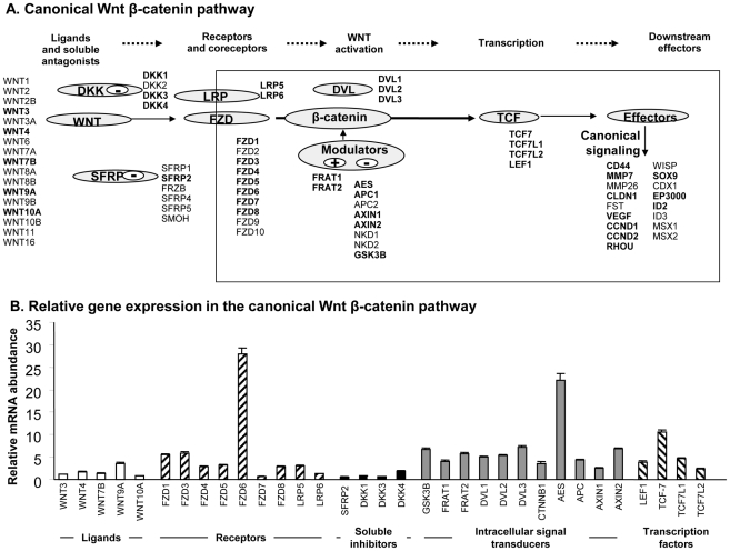
Background: The Wnt pathway mediates differentiation of epithelial tissues; depending on the tissue types, Wnt can either drive or inhibit the differentiation process. We hypothesized that key genes in the Wnt pathway are suppressed in the human airway epithelium under the stress of cigarette smoking, a stress associated with dysregulation of the epithelial differentiated state.

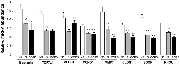
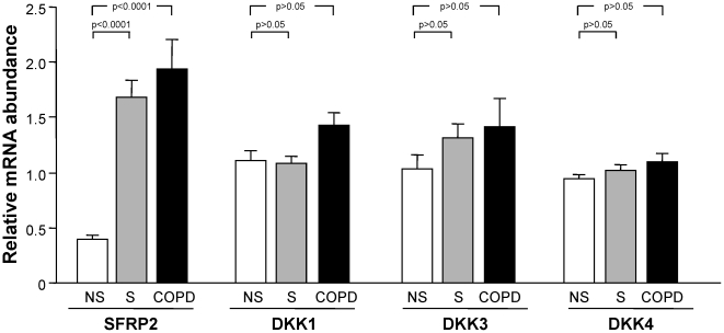
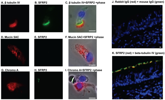
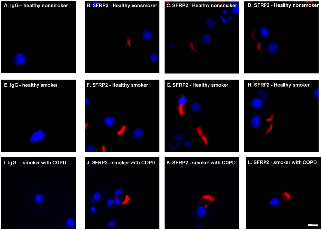
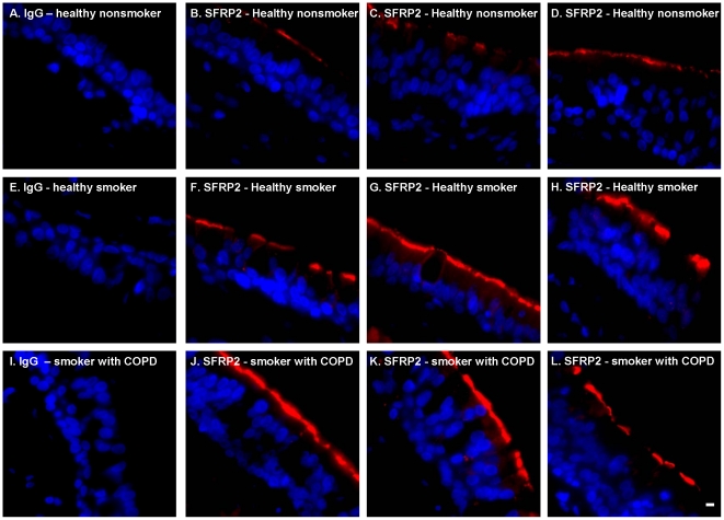
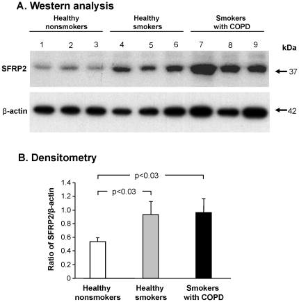
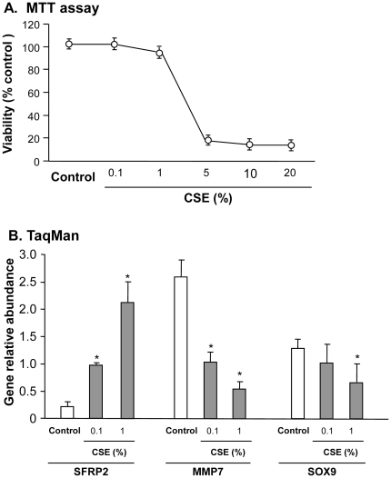
Methodology/principal findings: Microarrays were used to assess the expression of Wnt-related genes in the small airway epithelium (SAE) obtained via bronchoscopy and brushing of healthy nonsmokers, healthy smokers, and smokers with COPD. Thirty-three of 56 known Wnt-related genes were expressed in the SAE. Wnt pathway downstream mediators β-catenin and the transcription factor 7-like 1 were down-regulated in healthy smokers and smokers with COPD, as were many Wnt target genes. Among the extracellular regulators that suppress the Wnt pathway, secreted frizzled-related protein 2 (SFRP2), was up-regulated 4.3-fold in healthy smokers and 4.9-fold in COPD smokers, an observation confirmed by TaqMan Real-time PCR, Western analysis and immunohistochemistry. Finally, cigarette smoke extract mediated up-regulation of SFRP2 and down-regulation of Wnt target genes in airway epithelial cells in vitro.

Conclusions/significance: Smoking down-regulates the Wnt pathway in the human airway epithelium. In the context that Wnt pathway plays an important role in differentiation of epithelial tissues, the down-regulation of Wnt pathway may contribute to the dysregulation of airway epithelium differentiation observed in smoking-related airway disorders.


Conflict of interest statement

Competing Interests: The authors have declared that no competing interests exist.

Figures

Figure 1
Figure 1. The Wnt pathway expression in the small airway epithelium of human healthy nonsmokers.
A. Schematic of the pathway, showing which (in bold) ligands, soluble antagonists, receptors, co-receptors, intracellular activators, transcription factors and downstream effectors are expressed. B. Gene expression in canonical Wnt β-catenin pathway in healthy nonsmokers in small airway epithelium. Small airway epithelium from 47 normal nonsmokers were analyzed using the HG-U133 Plus 2.0 array and expression level normalized by chip only is plotted. Error bars represent the standard error.
Figure 2
Figure 2. Comparison of the relative expression of the Wnt pathway downstream and target genes in healthy nonsmokers (n = 47), healthy smokers (n = 58), and smokers with COPD (n = 22).
Analysis was carried out with HG-U133 plus 2.0 microarrays normalized by chip and gene. Each bar represents mean expression with standard error; * p<0.05 compared to healthy nonsmokers, * * p<0.01 compared to healthy nonsmokers.
Figure 3
Figure 3. Comparison of the relative expression of the Wnt pathway inhibitors in healthy nonsmokers (n = 47), healthy smokers (n = 58), and smokers with COPD (n = 22).
Analysis was carried out with HG-U133 plus 2.0 microarrays. Each bar represents mean expression with standard error; p values are represented in brackets above the bars.
Figure 4
Figure 4. Co-localization of SFRP2 and ciliated cell specific marker β-tubulin IV.
For Figures 4A–I , Cytospin preparations of airway epithelium from a healthy smoker were stained with antibodies against SFRP2 (green), β-tubulin IV (red), Mucin 5AC (red) and chromogranin A (red). A–C. Colocalization of SFRP2 and β-tubulin IV; D–F. Colocalization of SFRP2 and mucin 5AC; G–I. Colocalization of SFRP2 and chromogranin A. For Figure 4J–K , biopsies from a healthy smoker were stained with antibodies against SFRP2 (red) β- tubulin IV (green). J. IgG controls of SFRP2 and β-tubulin IV; and K. SFRP2 and β-tubulin IV co-localization.
Figure 5
Figure 5. Immunofluorescent assessment of SFRP2 expression in cytospin preparations of brushed small airway epithelium.
Small airway epithelial cell cytopreparations of healthy nonsmokers, healthy smokers and smokers with COPD were stained with anti-SFRP2 followed by a Cy3 conjugated secondary antibody (shown in red). Nuclei were stained with DAPI (shown in blue) A–D. Healthy nonsmokers. A. IgG control; B–D. Examples of anti-SFRP2. E–H. Healthy smokers. E. IgG control; F–H. Examples of anti-SFRP2. I–L. Smokers with COPD; I. IgG Control. J–L. Examples of anti-SFRP2. Bar = 10 µm.
Figure 6
Figure 6. Immunofluorescent assessment of SFRP2 expression in the endobronchial biopsies from large airway epithelium.
Endobronchial biopsies from large airway of healthy nonsmoker, healthy smokers and smokers with COPD were stained with anti-SFRP2 followed by a Cy3 conjugated secondary antibody (shown in red). Nuclei were stained with DAPI (shown in blue). A–D. Healthy nonsmokers. A. IgG control; B–D. Examples of anti-SFRP2. E–H. Healthy smokers. E. IgG control; F–H. Examples of anti-SFRP2. I–L. Smokers with COPD. I. IgG Control, J–L. Examples of anti-SFRP2. Bar = 10 µm.
Figure 7
Figure 7. Western analysis of SFRP2 protein expression in small airway epithelial cells.
A. Proteins were extracted from small airway epithelial cells of 3 healthy nonsmokers, 3 healthy smokers and 3 smokers with COPD. Shown is SFRP2 protein expression in healthy nonsmokers (lanes 1–3), healthy smokers (lanes 4–6) and smokers with COPD (lanes 7–9). Lower panel - same membrane probed with anti β-actin antibody, a control for protein loading. B. Quantification by densitometry of SFRP2 to β-actin. The ratio for SFRP2 to β-actin based on panel A is represented on the ordinate for the nonsmoker, smoker and smoker with COPD. Error bars represent the standard error.
Figure 8
Figure 8. In vitro cigarette smoke extract (CES) treatment of human 16HBE airway epithelial cells.
After 16HBE cells were exposed to different concentrations of CSE for 3 days, cell viability (MTT) and gene expression levels of Wnt signaling pathway inhibitor SFRP2 and target genes (MMP7, SOX9) were quantified using TaqMan real-time PCR. A. MTT assessment. B. Effect of CSE on gene expression of SFRP2, MMP7, SOX9 in 16HBE cells using 0.1 and 1% CSE concentrations that do not affect viability. SFRP2 gene expression was up-regulated when treated with both 0.1 and 1% CSE (4.6-fold and 10.1-fold, p<0.01 and p<0.001). Error bars represent the standard deviation. *p<0.05 compared to no CSE controls. For TaqMan real-time PCR, the average value of 0.1% CSE treatment group for each gene is determined as the calibrator.

Similar articles

Cited by

References

    1. Knight DA, Holgate ST. The airway epithelium: structural and functional properties in health and disease. Respirology. 2003;8:432–446. - PubMed
    1. Crystal RG, Randell SH, Engelhardt JF, Voynow J, Sunday ME. Airway epithelial cells: current concepts and challenges. Proc Am Thorac Soc. 2008;5:772–777. - PMC - PubMed
    1. Bishop AE. Pulmonary epithelial stem cells. Cell Prolif. 2004;37:89–96. - PMC - PubMed
    1. Rawlins EL, Ostrowski LE, Randell SH, Hogan BL. Lung development and repair: contribution of the ciliated lineage. Proc Natl Acad Sci U S A. 2007;104:410–417. - PMC - PubMed
    1. Stripp BR, Reynolds SD. Maintenance and repair of the bronchiolar epithelium. Proc Am Thorac Soc. 2008;5:328–333. - PMC - PubMed

Publication types

MeSH terms