Relationship between auditory thresholds, central spontaneous activity, and hair cell loss after acoustic trauma
- PMID: 21491427
- PMCID: PMC3140598
- DOI: 10.1002/cne.22644
Relationship between auditory thresholds, central spontaneous activity, and hair cell loss after acoustic trauma
Abstract
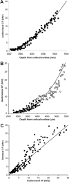
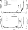
Acoustic trauma caused by exposure to a very loud sound increases spontaneous activity in central auditory structures such as the inferior colliculus. This hyperactivity has been suggested as a neural substrate for tinnitus, a phantom hearing sensation. In previous studies we have described a tentative link between the frequency region of hearing impairment and the corresponding tonotopic regions in the inferior colliculus showing hyperactivity. In this study we further investigated the relationship between cochlear compound action potential threshold loss, cochlear outer and inner hair cell loss, and central hyperactivity in inferior colliculus of guinea pigs. Two weeks after a 10-kHz pure tone acoustic trauma, a tight relationship was demonstrated between the frequency region of compound action potential threshold loss and frequency regions in the inferior colliculus showing hyperactivity. Extending the duration of the acoustic trauma from 1 to 2 hours did not result in significant increases in final cochlear threshold loss, but did result in a further increase of spontaneous firing rates in the inferior colliculus. Interestingly, hair cell loss was not present in the frequency regions where elevated cochlear thresholds and central hyperactivity were measured, suggesting that subtle changes in hair cell or primary afferent neural function are sufficient for central hyperactivity to be triggered and maintained.
Copyright © 2011 Wiley-Liss, Inc.
Figures


Similar articles
-
Hyperactivity in the auditory midbrain after acoustic trauma: dependence on cochlear activity.Neuroscience. 2009 Dec 1;164(2):733-46. doi: 10.1016/j.neuroscience.2009.08.036. Epub 2009 Aug 20. Neuroscience. 2009. PMID: 19699277
-
Effects of pulsatile electrical stimulation of the round window on central hyperactivity after cochlear trauma in guinea pig.Hear Res. 2016 May;335:128-137. doi: 10.1016/j.heares.2016.03.001. Epub 2016 Mar 10. Hear Res. 2016. PMID: 26970475
-
Influence of the paraflocculus on normal and abnormal spontaneous firing rates in the inferior colliculus.Hear Res. 2016 Mar;333:1-7. doi: 10.1016/j.heares.2015.12.021. Epub 2015 Dec 25. Hear Res. 2016. PMID: 26724754
-
Functional reorganization in chinchilla inferior colliculus associated with chronic and acute cochlear damage.Hear Res. 2002 Jun;168(1-2):238-49. doi: 10.1016/s0378-5955(02)00360-x. Hear Res. 2002. PMID: 12117524 Review.
-
Auditory plasticity and hyperactivity following cochlear damage.Hear Res. 2000 Sep;147(1-2):261-74. doi: 10.1016/s0378-5955(00)00136-2. Hear Res. 2000. PMID: 10962190 Review.
Cited by
-
Computational models of neurophysiological correlates of tinnitus.Front Syst Neurosci. 2012 May 8;6:34. doi: 10.3389/fnsys.2012.00034. eCollection 2012. Front Syst Neurosci. 2012. PMID: 22586377 Free PMC article.
-
Multisensory dysfunction accompanies crossmodal plasticity following adult hearing impairment.Neuroscience. 2012 Jul 12;214:136-48. doi: 10.1016/j.neuroscience.2012.04.001. Epub 2012 Apr 16. Neuroscience. 2012. PMID: 22516008 Free PMC article.
-
Using an appetitive operant conditioning paradigm to screen rats for tinnitus induced by intense sound exposure: Experimental considerations and interpretation.Front Neurosci. 2023 Feb 10;17:1001619. doi: 10.3389/fnins.2023.1001619. eCollection 2023. Front Neurosci. 2023. PMID: 36845432 Free PMC article.
-
Noise overexposure alters long-term somatosensory-auditory processing in the dorsal cochlear nucleus--possible basis for tinnitus-related hyperactivity?J Neurosci. 2012 Feb 1;32(5):1660-71. doi: 10.1523/JNEUROSCI.4608-11.2012. J Neurosci. 2012. PMID: 22302808 Free PMC article.
-
Cannabinoid CB1 Receptor Agonists Do Not Decrease, but may Increase Acoustic Trauma-Induced Tinnitus in Rats.Front Neurol. 2015 Mar 18;6:60. doi: 10.3389/fneur.2015.00060. eCollection 2015. Front Neurol. 2015. PMID: 25852639 Free PMC article.
References
-
- Atherley GR, Hempstock TI, Noble WG. Study of tinnitus induced temporarily by noise. J Acoust Soc Am. 1968;44(6):1503–1506. - PubMed
-
- Cody AR, Johnstone BM. Single auditory neuron response during acute acoustic trauma. Hear Res. 1980;3(1):3–16. - PubMed
-
- Cody AR, Johnstone BM. Acoustic trauma: single neuron basis for the “half-octave shift”. J Acoust Soc Am. 1981;70(3):707–711. - PubMed
Publication types
MeSH terms
Grants and funding
LinkOut - more resources
Full Text Sources

