. 2011 Sep 1;115(34):9590-602.
doi: 10.1021/jp200379m. Epub 2011 Apr 14.

Substantial contribution of the two imidazole rings of the His13-His14 dyad to Cu(II) binding in amyloid-β(1-16) at physiological pH and its significance

Affiliations

Substantial contribution of the two imidazole rings of the His13-His14 dyad to Cu(II) binding in amyloid-β(1-16) at physiological pH and its significance

Byong-kyu Shin et al. J Phys Chem A. .

Abstract

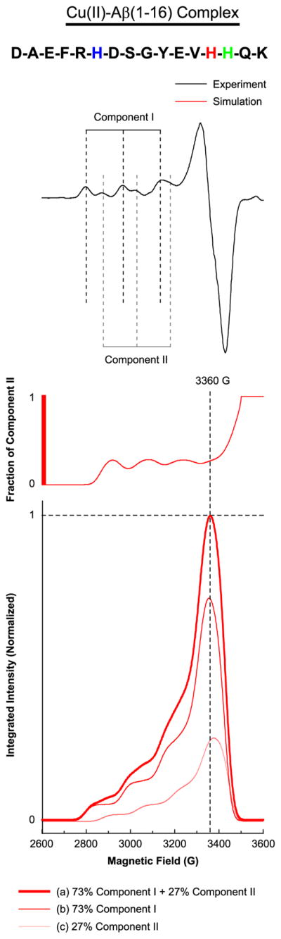
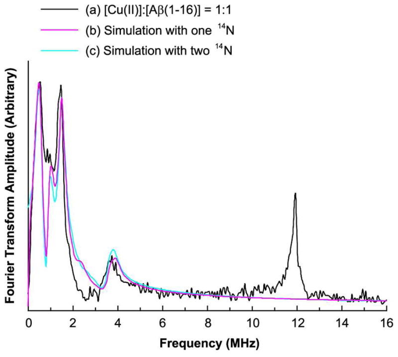
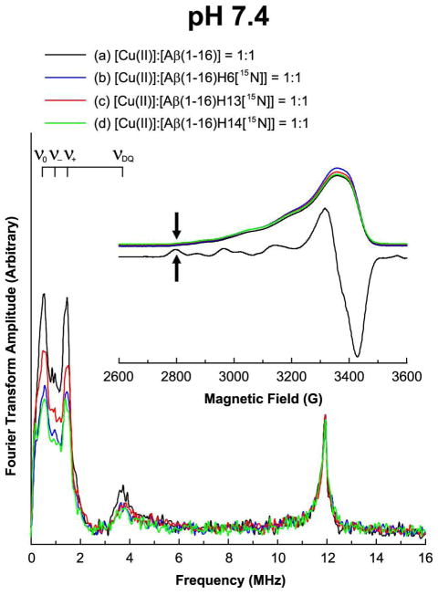
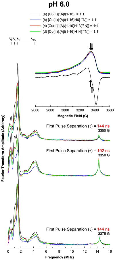
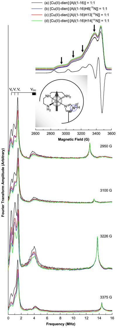
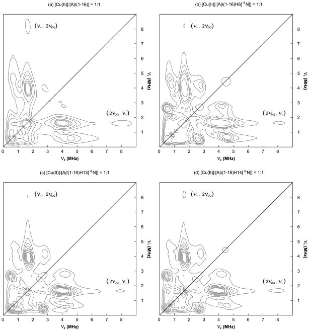
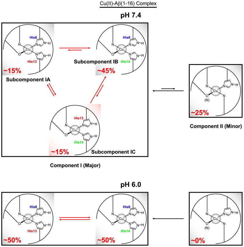
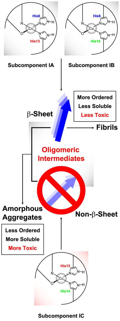
The interaction of amyloid-β (Aβ) peptide with Cu(II) appears to play an important role in the etiology of Alzheimer's disease. At physiological pH, the Cu(II) coordination in Aβ is heterogeneous, and there exist at least two binding modes in which Cu(II) is coordinated by histidine residues. Electron spin resonance studies have revealed a picture of the Cu(II) binding at a higher or lower pH, where only one of the two binding modes is almost exclusively present. We describe a procedure to directly examine the coordination of Cu(II) to each histidine residue in the dominant binding mode at physiological pH. We use nonlabeled and residue-specifically (15)N-labeled Aβ(1-16). For quantitative analysis, the intensities of three-pulse electron spin-echo envelope modulation (ESEEM) spectra are analyzed. Spectral simulations show that ESEEM intensities provide information about the contribution of each histidine residue. Indeed, the ESEEM experiments at pH 6.0 confirm the dominant contribution of His6 to the Cu(II) coordination as expected from the work of other researchers. Interestingly, however, the ESEEM data obtained at pH 7.4 reveal that the contributions of the three residues to the Cu(II) coordination are in the order of His14 ≈ His6 > His13 in the dominant binding mode. The order indicates a significant contribution from the simultaneous coordination by His13 and His14 at physiological pH, which has been underappreciated. These findings are supported by hyperfine sublevel correlation spectroscopy experiments. The simultaneous coordination by the two adjacent residues is likely to be present in a non-β-sheet structure. The coexistence of different secondary structures is possibly the molecular origin for the formation of amorphous aggregates rather than fibrils at relatively high concentrations of Cu(II). Through our approach, precise and useful information about Cu(II) binding in Aβ(1-16) at physiological pH is obtained without any side-chain modification, amino acid residue replacement, or pH change, each of which might lead to an alteration in the peptide structure or the coordination environment.


Figures

Figure 1
Figure 1
Multiple components present in the Cu(II)–Aβ(1–16) complex suggested by the experimental and simulated CW-ESR spectrum of Aβ(1–16) mixed with an equimolar amount of Cu(II). The amino acid sequence of Aβ(1–16) is illustrated with His6, His13, and His14 indicated in blue, red, and green, respectively. The experimentally obtained CW-ESR spectrum, which contains two clearly distinguished components, Component I and Component II, is shown at the top. The fraction of Component II as a function of the magnetic field is shown at the middle. At the bottom, the curves corresponding to 73% Component I, 27% Component II, and the mixture thereof are shown in red, pink, and bold red, respectively. Component II accounts for approximately a quarter of the Cu(II)–Aβ(1–16) complex at 3360 G while there is practically no contribution of Component II below 2810 G.
Figure 2
Figure 2
Experimentally obtained and simulated three-pulse ESEEM spectra of the isotopically nonlabeled Aβ(1–16) peptide mixed with an equimolar amount of Cu(II) at 2800 G at pH 7.4. The Simulated spectra with two ESEEM-active 14N nuclei are in good agreement with the experimental result.
Figure 3
Figure 3
Three-pulse ESEEM and field-swept echo detected spectra of the nonlabeled and 15N-labeled Aβ(1–16) analogues mixed with an equimolar amount of Cu(II) at 2800 G at pH 7.4. The decrease in the 14N-ESEEM intensity below 8 MHz is more prominent when His6 or His14 is enriched with 15N. On the other hand, the 1H-ESEEM intensity of each spectrum is essentially identical.
Figure 4
Figure 4
Three-pulse ESEEM and field-swept echo detected spectra of the nonlabeled and 15N-labeled Aβ(1–16) analogues mixed with an equimolar amount of Cu(II) at pH 6.0. The decrease in the 14N-ESEEM intensity below 8 MHz is more prominent when His6 is enriched with 15N. On the other hand, the 1H-ESEEM intensity of each spectrum is essentially identical.
Figure 5
Figure 5
Three-pulse ESEEM and field-swept echo detected spectra of the nonlabeled and 15N-labeled Aβ(1–16) analogues mixed with an equimolar amount of the Cu(II)–dien complex at pH 7.4. The CW-ESR spectrum of the nonlabeled version at pH 7.4 is also presented. The CW-ESR spectrum and the field-swept echo detected spectra display characteristic features of dien–Cu(II)–imidazole ternary complexes. The decrease in the 14N-ESEEM intensity below 8 MHz is most prominent when His14 is enriched with 15N. On the other hand, the 1H-ESEEM intensity is not significantly affected by the replacement of 14N with 15N.
Figure 6
Figure 6
14N- and 15N-ESEEM regions of the HYSCORE spectra of the nonlabeled and 15N-labeled Aβ(1–16) analogues mixed with an equimolar amount of Cu(II) at 3360 G at pH 7.4. Each of the four spectra has a cross-peak around (1.6 MHz, 8.0 MHz), which indicates multiple histidine coordination.
Figure 7
Figure 7
Difference in Cu(II)-binding modes of Aβ(1–16) between pH 7.4 and pH 6.0 suggested by ESR spectroscopy. While His6 accounts for approximately 50% of the Cu(II)–histidine coordination in Component I at pH 6.0, the contribution of His14 is at least as significant as that of His6 at pH 7.4. At pH 7.4, Component I is composed of three subcomponents, Subcomponent IA, IB, and IC, in each of which two imidazole rings from two different histidine residues coordinate to Cu(II).
Figure 8
Figure 8
Formation of fibrils and amorphous aggregates of Aβ peptide in the presence of Cu(II) and the suggested role of the simultaneous intramolecular coordination by two imidazole rings of His13 and His14. With a significant amount of Subcomponent IC, the β-sheet and non–β-sheet structures coexist, which leads to the preference of amorphous aggregates over fibrils.

Similar articles

Cited by

References

    1. Hardy J, Selkoe DJ. Science. 2002;297:353–356. - PubMed
    1. Kirkitadze MD, Bitan G, Teplow DB. J Neurosci Res. 2002;69:567–577. - PubMed
    1. Carrotta R, Manno M, Bulone D, Martorana V, Biagio PLS. J Biol Chem. 2005;280:30001–30008. - PubMed
    1. Haass C, Selkoe DJ. Nat Rev Mol Cell Biol. 2007;8:101–112. - PubMed
    1. Crouch PJ, Harding SME, White AR, Camakaris J, Bush AI, Masters CL. Int J Biochem Cell Biol. 2008;40:181–198. - PubMed

Publication types

MeSH terms