. 2011 Nov;17(4):387-97.
doi: 10.1111/j.1600-0846.2011.00508.x. Epub 2011 Apr 15.

Characterization and quantification of wound-induced hair follicle neogenesis using in vivo confocal scanning laser microscopy

Affiliations

Characterization and quantification of wound-induced hair follicle neogenesis using in vivo confocal scanning laser microscopy

Chengxiang Fan et al. Skin Res Technol. 2011 Nov.

Abstract

Background: In vivo confocal scanning laser microscopy (CSLM) is a recently developed non-invasive technique for visualizing microscopic structures with the skin. CSLM has been used to characterize proliferative and inflammatory skin diseases, neoplastic skin lesions and pigmented lesions.

Objective: Here, we assessed the ability of CSLM to evaluate the formation of neogenic hair follicles after a full-thickness wound in mice.

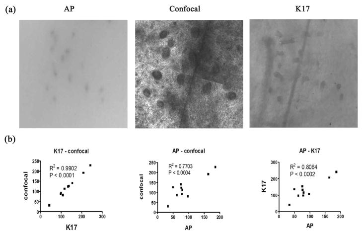
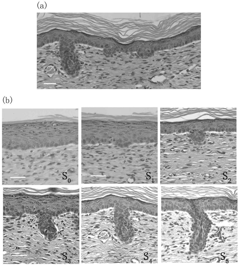
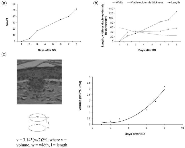
Methods: Full-thickness wounds were made on the dorsal skin of 3-week-old mice. After scab detachment (SD), the number, width, length, space and volume of neogenic hair follicles were analyzed using CSLM. The results were compared with those from conventional methods, including staining for alkaline phosphatase (AP) and keratin 17 (K17) as well as histology.

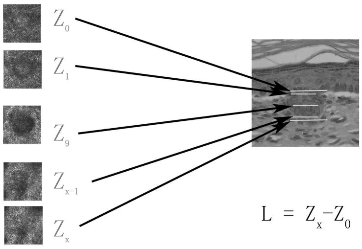
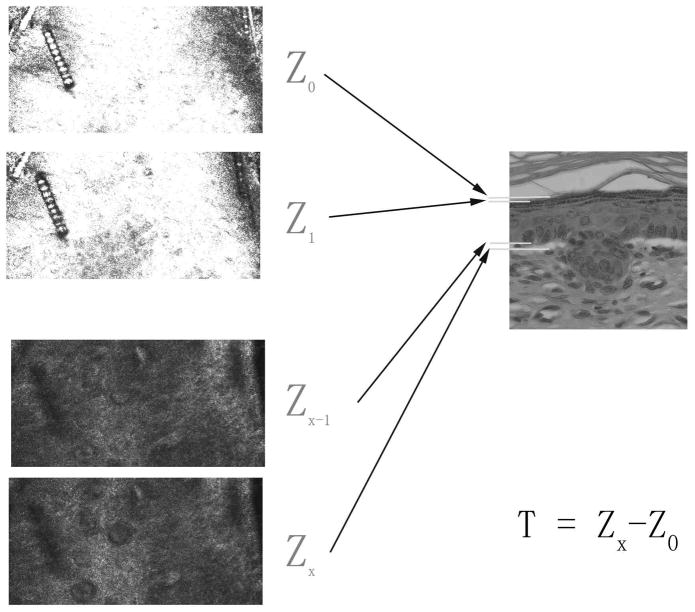
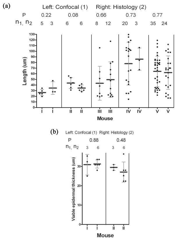
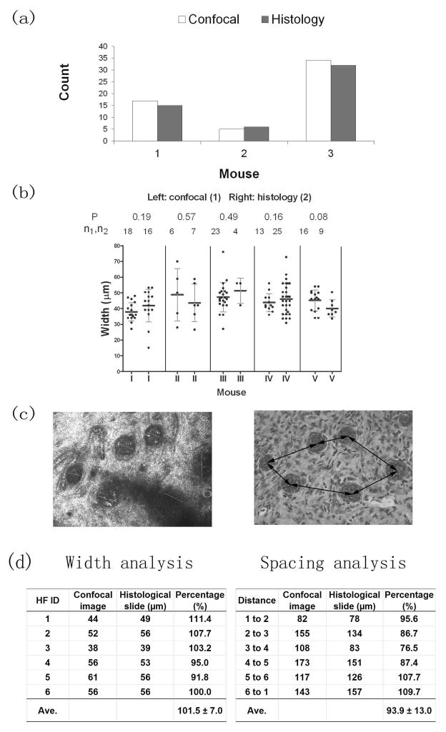
Results: Quantification of neogenic hair follicles using CSLM compared favorably with the results from direct measurements on isolated epidermal tissue after immunostaining for K17, a marker for the epithelial portion of new hair follicles. CSLM detected 89% of K17-stained follicles. CSLM more accurately quantified the number of new follicles compared with AP staining, which detects the dermal portion of the new follicle. The width and length measurement from CSLM and histology were very close and correlated with each other. The minimum length of a neogenic hair follicle that could be detected by CSLM was 21 μm. The space between neogenic hair follicles was decreased in histological sections compared with CSLM.

Conclusion: CSLM is an accurate and valuable method for counting and measuring neogenic hair follicles non-invasively. CSLM produces images similar to histology in mice. Measurements of microstructures using CSLM more accurately reflect actual sizes as this technique avoids fixation artifacts. In vivo visualization of developing follicles with CSLM allows the detection of serial changes in hair follicle formation, thus conserving the numbers of mice required for studies and improving the detection of temporal changes in developing hair follicles.


Figures

Figure 1
Figure 1. Length measurement from CSLM image analysis
Sequential stack images from SC to dermis were analyzed. Z0 is defined as the deepest layer where the basal cells are observed, Z1 is defined as the layer where the hair follicle germs are seen clearly, Zx−1 is the deepest layer where germs can still be seen and (Zx) is defined as the layer where the germs are no longer observed. The depth information of these layers was obtained by normalizing the depth information relative to the stratum corneum surface and using the calibrated image steps from the skin surface. Once Z0 and Zx were determined,the length of the follicle (L) was calculated from the difference between these two layers.
Figure 2
Figure 2. Thickness measurement of viable epidermis from CSLM image analysis
Sequential stack images from SC to dermis were analyzed. Z0 is defined as the deepest layer where keratinocytes have not yet appeared, Z1 is the layer where keratinocytes begin to appear, Zx−1 is defined as the last layer where basal cells are observed and Zx is defined as the layer where the basal cells are no longer observed. The difference in depth between Zx and Z0 is defined as the thickness of viable epidermis (T).
Figure 3
Figure 3. Correlation of neogenic hair follicle counts between AP, confocal and K17 assays
Fig. 3a) A representative match of AP, K17, and CSLM images used to quantify neogenic hair follicles. Fig. 3b) Demonstrates correlation analysis for the total counts of neogenic hair follicles between these assays.
Figure 4
Figure 4. Different stages of neogenic hair follicles in the hair follicle neogenesis following wounding
Fig. 4a) shows the asynchrony of neogenic hair follicles. Different stages of neogenic hair follicles are seen in one sample after SD. Fig. 4b) shows representative neogenic hair follicles of different stages (S0–S5) seen at various times after SD. S2 is the earliest germ detectable with CSLM. Scale bar: 25 μm
Figure 5
Figure 5. Comparison of length and viable epidermal thickness measurements between CSLM and histological methods
Fig 5a) shows the comparison of length measurement between CSLM and histology methods. Fig 5b) shows the results of viable epidermal thickness measurement from both methods. Measurements n1 and n2 are determined from confocal and histology methods respectively. P values are representative of the t-test of measurements from these two methods.
Figure 6
Figure 6. Comparison of width and space measurements between CSLM and histological assays
Fig 6a) Total hair germ counts from confocal and histological assays. Fig. 6b) Comparison of width measurements between confocal and histological assays. Measurements n1 and n2 are from confocal and histology methods respectively. P values are representative of the t-test of measurements from these two methods. Fig. 6c) Is a representative match between CSLM and histological images. The confocal image (on left) also shows how width measurement was made, whereas the right histological image shows the space measurement. Fig 6d) shows the width and space analysis in the above pair of images.
Figure 7
Figure 7. Development of neogenic hair follicles over time
The same mouse was imaged with CSLM from Day 1 to Day 8 after SD. The same regions were imaged each day. Figs. 7a, b, c, d, e and f show the wound areas of Day 1, 2, 3, 6, 7 and 8 after SD respectively.
Figure 8
Figure 8. Dynamic changes of the parameters of neogenic hair follicles after SD
A mouse was imaged with CSLM from Day 1 through Day 8 after SD. Fig. 8a) demonstrates the dynamic change in the number of neogenic hair follicles. Fig. 8b) shows the dynamic changes in the length and width of neogenic hair follicles and thickness of viable epidermis. Fig 8c) Demonstrates the volume dynamics of a model neogenic hair follicle.

References

    1. Billingham RE, Russell PS. Incomplete wound contracture and the phenomenon of hair neogenesis in rabbits' skin. Nature. 1956 Apr 28;177(4513):791–2. - PubMed
    1. Breedis C. Regeneration of hair follicles and sebaceous glands from the epithelium of scars in the rabbit. Cancer Res. 1954 Sep;14(8):575–9. - PubMed
    1. Lacassagne A, Latarjet R. Action of methylcholanthrene on certain scars of the skin in mice. Cancer Res. 1946;(6):183–8. - PubMed
    1. Kligman AM, Strauss JS. The formation of vellus hair follicles from human adult epidermis. J Invest Dermatol. 1956 Jul;27(1):19–23. - PubMed
    1. Ito M, Yang Z, Andl T, Cui C, Kim N, Millar SE, et al. Wnt-dependent de novo hair follicle regeneration in adult mouse skin after wounding. Nature. 2007 May 17;447(7142):316–20. - PubMed

Publication types

MeSH terms

LinkOut - more resources