

Inhibition of nuclear factor kappa-B signaling reduces growth in medulloblastoma in vivo
- PMID: 21492457
- PMCID: PMC3094324
- DOI: 10.1186/1471-2407-11-136
Inhibition of nuclear factor kappa-B signaling reduces growth in medulloblastoma in vivo
Abstract
Background: Medulloblastoma is a highly malignant pediatric brain tumor that requires surgery, whole brain and spine irradiation, and intense chemotherapy for treatment. A more sophisticated understanding of the pathophysiology of medulloblastoma is needed to successfully reduce the intensity of treatment and improve outcomes. Nuclear factor kappa-B (NFκB) is a signaling pathway that controls transcriptional activation of genes important for tight regulation of many cellular processes and is aberrantly expressed in many types of cancer.
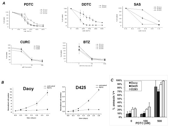
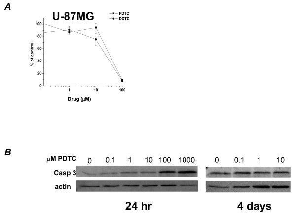
Methods: To test the importance of NFκB to medulloblastoma cell growth, the effects of multiple drugs that inhibit NFκB, pyrrolidine dithiocarbamate, diethyldithiocarbamate, sulfasalazine, curcumin and bortezomib, were studied in medulloblastoma cell lines compared to a malignant glioma cell line and normal neurons. Expression of endogenous NFκB was investigated in cultured cells, xenograft flank tumors, and primary human tumor samples. A dominant negative construct for the endogenous inhibitor of NFκB, IκB, was prepared from medulloblastoma cell lines and flank tumors were established to allow specific pathway inhibition.
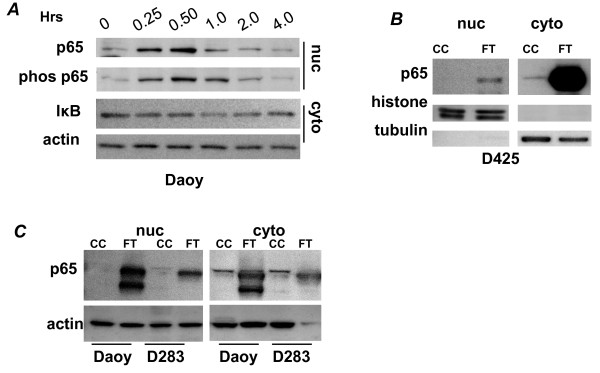
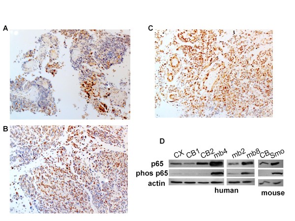
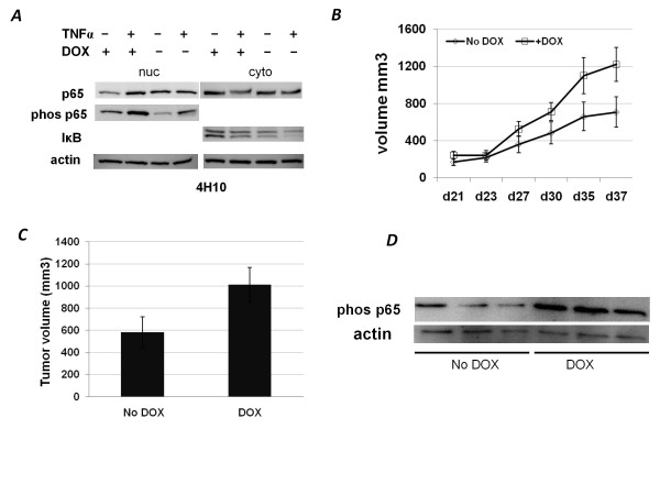
Results: We report high constitutive activity of the canonical NFκB pathway, as seen by Western analysis of the NFκB subunit p65, in medulloblastoma tumors compared to normal brain. The p65 subunit of NFκB is extremely highly expressed in xenograft tumors from human medulloblastoma cell lines; though, conversely, the same cells in culture have minimal expression without specific stimulation. We demonstrate that pharmacological inhibition of NFκB in cell lines halts proliferation and leads to apoptosis. We show by immunohistochemical stain that phosphorylated p65 is found in the majority of primary tumor cells examined. Finally, expression of a dominant negative form of the endogenous inhibitor of NFκB, dnIκB, resulted in poor xenograft tumor growth, with average tumor volumes 40% smaller than controls.
Conclusions: These data collectively demonstrate that NFκB signaling is important for medulloblastoma tumor growth, and that inhibition can reduce tumor size and viability in vivo. We discuss the implications of NFκB signaling on the approach to managing patients with medulloblastoma in order to improve clinical outcomes.
Figures
Similar articles
-
Bortezomib induces apoptosis and growth suppression in human medulloblastoma cells, associated with inhibition of AKT and NF-ĸB signaling, and synergizes with an ERK inhibitor.Cancer Biol Ther. 2012 Apr;13(6):349-57. doi: 10.4161/cbt.19239. Epub 2012 Apr 1. Cancer Biol Ther. 2012. PMID: 22313636 Free PMC article.
-
C086, a novel analog of curcumin, induces growth inhibition and down-regulation of NFκB in colon cancer cells and xenograft tumors.Cancer Biol Ther. 2011 Nov 1;12(9):797-807. doi: 10.4161/cbt.12.9.17671. Epub 2011 Nov 1. Cancer Biol Ther. 2011. PMID: 21900746
-
An anti-leishmanial thiadiazine agent induces multiple myeloma cell apoptosis by suppressing the nuclear factor kappaB signalling pathway.Br J Cancer. 2014 Jan 7;110(1):63-70. doi: 10.1038/bjc.2013.711. Epub 2013 Nov 14. Br J Cancer. 2014. PMID: 24231956 Free PMC article.
-
Medulloblastoma: from molecular pathology to therapy.Clin Cancer Res. 2008 Feb 15;14(4):971-6. doi: 10.1158/1078-0432.CCR-07-2072. Clin Cancer Res. 2008. PMID: 18281528 Free PMC article. Review.
-
Exploring the role and clinical implications of proteasome inhibition in medulloblastoma.Pediatr Blood Cancer. 2021 Oct;68(10):e29168. doi: 10.1002/pbc.29168. Epub 2021 Jun 11. Pediatr Blood Cancer. 2021. PMID: 34114315 Free PMC article. Review.
Cited by
-
Transcription Factors and Methods for the Pharmacological Correction of Their Activity.Int J Mol Sci. 2025 Jul 2;26(13):6394. doi: 10.3390/ijms26136394. Int J Mol Sci. 2025. PMID: 40650173 Free PMC article. Review.
-
Combined MEK and JAK/STAT3 pathway inhibition effectively decreases SHH medulloblastoma tumor progression.Commun Biol. 2022 Jul 14;5(1):697. doi: 10.1038/s42003-022-03654-9. Commun Biol. 2022. PMID: 35835937 Free PMC article.
-
The role of the ubiquitin proteasome system in cerebellar development and medulloblastoma.Mol Brain. 2015 Oct 17;8(1):64. doi: 10.1186/s13041-015-0155-5. Mol Brain. 2015. PMID: 26475605 Free PMC article. Review.
-
The beneficial effects of curcumin on aging and age-related diseases: from oxidative stress to antioxidant mechanisms, brain health and apoptosis.Front Aging Neurosci. 2025 Jan 20;17:1533963. doi: 10.3389/fnagi.2025.1533963. eCollection 2025. Front Aging Neurosci. 2025. PMID: 39906716 Free PMC article. Review.
-
Diethyldithiocarbamate induces apoptosis in HHV-8-infected primary effusion lymphoma cells via inhibition of the NF-κB pathway.Int J Oncol. 2012 Apr;40(4):1071-8. doi: 10.3892/ijo.2011.1313. Epub 2011 Dec 20. Int J Oncol. 2012. PMID: 22200846 Free PMC article.
References
-
- Pollack IF. In: Tumors of the pediatric central nervous system. Keating RF, Goodrich JT, Packer RJ, editor. New York: Thieme Medical Publishers, Inc; 2001. Infratentorial primitive neuroectodermal tumors; pp. 251–264.
-
- Hatton BA, Villavicencio EH, Tsuchiya KD, Pritchard JI, Ditzler S, Pullar B, Hansen S, Knoblaugh SE, Lee D, Eberhart CG, Hallahan AR, Olson JM. The Smo/Smo model: hedgehog-induced medulloblastoma with 90% incidence and leptomeningeal spread. Cancer Res. 2008;68:1768–1776. doi: 10.1158/0008-5472.CAN-07-5092. - DOI - PubMed
Publication types
MeSH terms
Substances
LinkOut - more resources
Full Text Sources
Other Literature Sources
Molecular Biology Databases
