. 2011 Jun 5;414(2):153-63.
doi: 10.1016/j.virol.2011.03.019. Epub 2011 Apr 13.

Persistence of viral DNA in the epithelial basal layer suggests a model for papillomavirus latency following immune regression

Affiliations

Persistence of viral DNA in the epithelial basal layer suggests a model for papillomavirus latency following immune regression

Gareth Adam Maglennon et al. Virology. .

Abstract

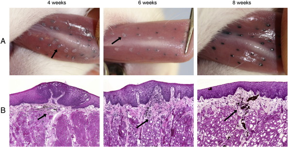
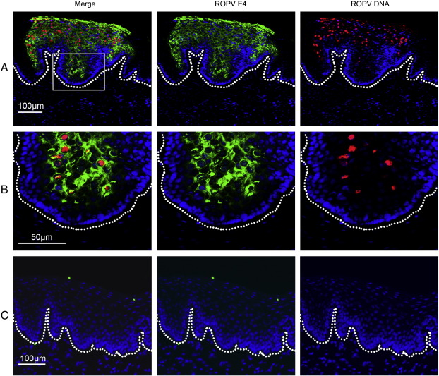
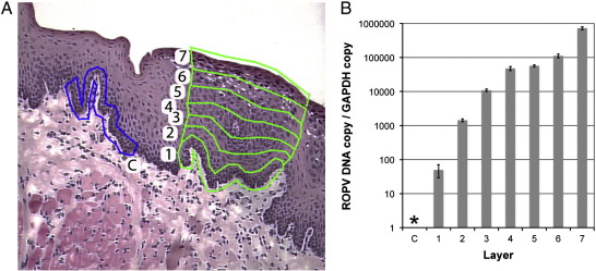
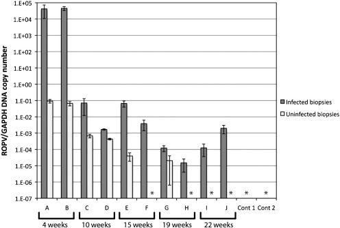
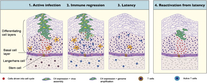
Rabbit oral papillomavirus (ROPV) causes benign and spontaneously regressing oral lesions in rabbits, and is a useful model of disease associated with low-risk human papillomavirus types. Here we have adapted the ROPV system to study papillomavirus latency. Following lesion regression, ROPV DNA persists at the majority of regressed sites at levels substantially lower than those found in productive papillomas. Spliced viral transcripts were also detected. ROPV persistence in the absence of disease could be demonstrated for a year following infection and lesion-regression. This was not associated with completion of the virus life-cycle or new virion production, indicating that ROPV persists in a latent state. Using novel laser capture microdissection techniques, we could show that the site of latency is a subset of basal epithelial cells at sites of previous experimental infection. We hypothesize that these cells are epithelial stem cells and that reactivation of latency may be a source of recurrent disease.


Figures

Fig. 1
Fig. 1
Formation of papillomas following experimental infection. The mucosa of the ventral surface of the tongue was infected using a crude papilloma homogenate mixed with tattoo ink. The gross (row A) and histological (row B) appearance of lesions is shown at 4, 6 and 8 weeks post-infection by photographs taken under general anaesthesia and H&E stained cryosections obtained after culling of animals. The locations of tattoo ink on H&E stained sections are shown by arrows.
Fig. 2
Fig. 2
Fluorescent in situ hybridization of ROPV DNA. Presence of ROPV DNA was determined in tissue sections from experimentally infected rabbits using a full-length DIG-labeled probe. Sections were also stained for the presence of ROPV E1^E4 protein. The basal layer of epithelial cells is marked by a dotted line. Fluorescent images are shown for a productive papilloma at 4 weeks post-infection at ×10 (row A) and ×20 (row B). Detection of ROPV DNA generally coincides with expression of E1^E4 protein. ROPV DNA is not visible in the basal layers of the epithelium. At 8 weeks post-infection (row C) following the regression of papillomas, ROPV DNA and E1^E4 protein are no longer visible.
Fig. 3
Fig. 3
Detection of ROPV in papillomas using LCM. LCM was used to dissect seven (1 to 7) sequential layers of epithelial cells from papillomas harvested at 4 weeks post-infection (A). Uninfected adjacent control samples (layer C) were also obtained. DNA was extracted and real-time PCR performed for quantification of ROPV DNA and GAPDH reference gene. The graph in image B shows a ratio of ROPV copy to GAPDH copy number (with standard error) for each corresponding layer shown in image A. ROPV DNA was not detected in basal layer samples (layer C) taken immediately adjacent to papillomas (*). ROPV DNA was always detected in basal layer samples obtained from papillomas (layer 1). A gradual rise in ROPV DNA copy number was seen between the basal layers and the epithelial surface within papillomas. The degree of amplification was approximately four to five log-fold.
Fig. 4
Fig. 4
ROPV DNA copy number at infected and uninfected sites. Pairs of rabbits infected with ROPV were culled at successive intervals. ROPV DNA copy number was determined relative to GAPDH at five experimentally infected and five uninfected sites after culling. The average ratio of ROPV DNA to GAPDH reference gene is shown with standard error. Samples marked * indicates that no ROPV DNA was detected in any of the five samples. Two control rabbits underwent scarification but were not infected with ROPV. ROPV DNA was not detected at any of the scarified or non-scarified sites.
Fig. 5
Fig. 5
Collection of LCM samples from tissue sections. Sites of experimental infection were identified by means of tattoo marks (arrowheads) on tissue H&E stained tissue sections. The following tissue samples were selected for dissection using LCM (A): blank slide membrane (blue), stromal tissue (yellow), uninfected adjacent basal epithelial cells (green) and basal epithelial cells from immediately above the tattoo mark (red). Tissue samples were collected using LCM into microcentrifuge tubes (B).
Fig. 6
Fig. 6
Real-time PCR detection of ROPV DNA and RNA transcripts. ROPV DNA copy number was assessed by real-time PCR relative to GAPDH reference gene in eleven ROPV-infected rabbits. The average ratio of ROPV to GAPDH copy number from five tongue tissue samples is shown with standard error (A). Levels of E1^E4 transcript were also assessed in single tissue samples for each rabbit. Samples marked by * had no ROPV DNA and/or E1^E4 transcripts detected. During latency, E1^E4 spliced transcripts were detected in five rabbits (N, O, P, Q & U). Further RNA analysis was performed on tissues from these rabbits to detect and quantify transcripts originating from the E1, E2, E6 and E7 open reading frames (B). No-RT controls were run for all samples and were always negative (marked *). In addition to detecting spliced E1^E4 ROPV transcripts, RNA sequences originating from the E1, E2, E6 and E7 open reading frames were detected in all of these five rabbits.
Fig. 7
Fig. 7
Expression of E1^E4, L1 and MCM7 proteins in ROPV papillomas and latency. Tissue sections were obtained from ROPV papillomas and following regression of lesions during latency. Detection of MCM7, E1^E4 and L1 proteins was performed by immunofluorescence. In ROPV papillomas, expression of MCM7 was present from the basal layer of the epithelium to the mid layers, consistent with expression of E7 protein and maintenance of cells in a proliferative state (panel A). Expression of E1^E4 protein started in the mid layers of the epithelium and overlapped for one or two cell layers with MCM7. Expression was present to the surface of the epithelium and was primarily cytoplasmic in location. Expression of L1 capsid protein was observed in the upper layers of the epithelium consistent with new virion formation and was nuclear in location. Following the regression of papillomas, an extensive analysis of tissue sections failed to demonstrate the presence of E1^E4 and L1 viral proteins (panel B). MCM7 expression was still present in a subpopulation of basal epithelial cells only (panel B) and mirrored that of normal uninfected epithelium (panel C).
Fig. 8
Fig. 8
A model of papillomavirus latency following immune regression. An active infection follows entry of papillomavirus into an epithelial stem cell in the basal layer of the epithelium. Cells in the basal layer and above are driven into cell cycle allowing genome amplification and new virion production to occur in the intermediate and upper cell layers. Viral strategies, such as low level protein production in the lower epithelial layers, assist the virus in evading immune detection. Triggering of an effective immune response leads to immune regression, accompanied by infiltration of predominantly T cells. Viral gene expression is shut off and lesion regression occurs. Viral latency may ensue with viral genomes restricted to stem cells in the basal layer of the epithelium. Reactivation of latency is prevented by host immune surveillance. Factors such as immune suppression may allow reactivation to occur. Completion of the virus life cycle may or may not be associated with reappearance of a visible lesion.

Similar articles

Cited by

References

    1. Abramson A.L., Nouri M., Mullooly V., Fisch G., Steinberg B.M. Latent human papillomavirus infection is comparable in the larynx and trachea. J. Med. Virol. 2004;72(3):473–477. - PubMed
    1. Amella C.A., Lofgren L.A., Ronn A.M., Nouri M., Shikowitz M.J., Steinberg B.M. Latent infection induced with cottontail rabbit papillomavirus, A model for human papillomavirus latency. Am. J. Pathol. 1994;144(6):1167–1171. - PMC - PubMed
    1. Broker T.R., Jin, Croom-Rivers A., Bragg S.M., Richardson M., Chow L.T., Vermund S.H., Alvarez R.D., Pappas P.G., Squires K.E., Hoesley C.J. Viral latency—the papillomavirus model. Dev Biol (Basel) 2001;106:443–451. discussion 452–3, 465–75. - PubMed
    1. Christensen N.D., Cladel N.M., Reed C.A., Han R. Rabbit oral papillomavirus complete genome sequence and immunity following genital infection. Virology. 2000;269(2):451–461. - PubMed
    1. Embers M.E., Budgeon L.R., Pickel M., Christensen N.D. Protective immunity to rabbit oral and cutaneous papillomaviruses by immunization with short peptides of L2, the minor capsid protein. J. Virol. 2002;76(19):9798–9805. - PMC - PubMed

Publication types

MeSH terms