. 2011 Jun 3;286(22):19410-6.
doi: 10.1074/jbc.M110.205211. Epub 2011 Apr 14.

Physical and functional interaction of the HECT ubiquitin-protein ligases E6AP and HERC2

Affiliations

Physical and functional interaction of the HECT ubiquitin-protein ligases E6AP and HERC2

Simone Kühnle et al. J Biol Chem. .

Abstract

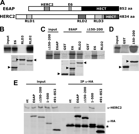
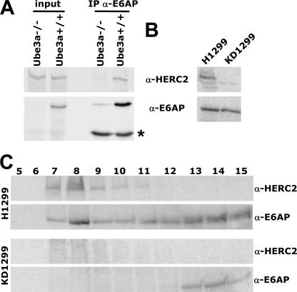
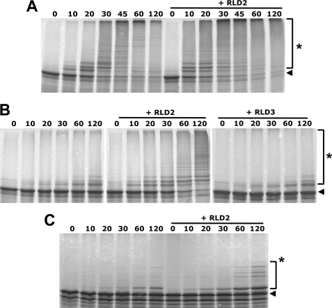
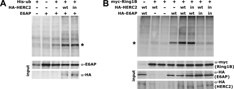
Deregulation of the ubiquitin-protein ligase E6AP contributes to the development of the Angelman syndrome and to cervical carcinogenesis suggesting that the activity of E6AP needs to be under tight control. However, how E6AP activity is regulated at the post-translational level under non-pathologic conditions is poorly understood. In this study, we report that the giant protein HERC2, which is like E6AP a member of the HECT family of ubiquitin-protein ligases, binds to E6AP. The interaction is mediated by the RCC1-like domain 2 of HERC2 and a region spanning amino acid residues 150-200 of E6AP. Furthermore, we provide evidence that HERC2 stimulates the ubiquitin-protein ligase activity of E6AP in vitro and within cells and that this stimulatory effect does not depend on the ubiquitin-protein ligase activity of HERC2. Thus, the data obtained indicate that HERC2 acts as a regulator of E6AP.


Figures

FIGURE 1.
FIGURE 1.
The HECT ubiquitin ligase HERC2 binds to a region within the N terminus of E6AP. A, schematic structure of human E6AP (isoform 1) and human HERC2. For E6AP, the position of the HERC2 and E6 binding domains (residues 150–200 and 378–395, respectively) and the HECT domain (residues 492–852) are indicated. For HERC2, the positions of RLD1, RLD2, and RLD3 (residues 513–778, 2958–3326, and 3951–4318, respectively) and the HECT domain (residues 4457–4834) are indicated. B, RLD2, RLD3, and the HPV16 E6 oncoprotein were bacterially expressed as GST fusion proteins and purified by affinity chromatography using glutathione-Sepharose. Similar amounts of the various fusion proteins (lower panel; running positions of the respective full-length proteins are indicated by arrowheads) were incubated with in vitro translated radiolabeled E6AP (isoform 1). After 3 h at 4 °C, glutathione beads were centrifuged, supernatants were removed, and beads were washed. The amount of E6AP bound to the various GST fusion proteins was determined by SDS-PAGE followed by fluorography (upper panel). input, corresponds to 20% of in vitro translated E6AP used for binding reactions. C, binding reactions and analysis of bound proteins (upper panel) were performed as in B with GST fusion proteins of RLD2 and HPV16 E6 as affinity matrix (for amounts, see the lower panel; running positions of the respective full-length proteins are indicated by arrowheads) and in vitro translated radiolabeled E6AP or E6APΔ150–200 (deletion of amino acid residues 150–200) as indicated. input, corresponds to 20% of in vitro translated E6AP and E6APΔ150–200 used for binding reactions. D, binding reactions and analysis of bound proteins were performed as in B with a GST fusion protein of amino acid residues 150–200 of E6AP (150–200) and in vitro translated radiolabeled RLD2 (upper panel). GST was used as an affinity matrix control. input, corresponds to 20% of in vitro translated RLD2 used for binding reactions. For amounts of GST and GST-E6AP150–200 used as affinity matrix, see the lower panel. E, H1299 cells were transfected with expression constructs for the HA-tagged forms of E6AP indicated. Protein extracts were prepared 24 h after transfection. 20% of the respective extracts were used to determine levels of the various forms of E6AP and endogenous HERC2 by Western blot analysis as indicated (input). To the remainder of the extracts, an anti-HA antibody was added, mixtures were incubated for 4 h at 4 °C, and bound proteins were precipitated by Protein A-Sepharose. Precipitated proteins were detected by Western blot analysis as indicated. nt, untransfected cell extracts were used as control. The band indicated with an star represents the heavy chain of the anti-HA antibody used.
FIGURE 2.
FIGURE 2.
Copurification of endogenous E6AP and HERC2. A, extracts were prepared from MEFs derived from E6AP null embryos (Ube3a−/−) and matched littermates (Ube3a+/+). Levels of E6AP and HERC2 were either directly analyzed by Western blot analysis (input) or extracts were first subjected to immunoprecipitation with an anti-E6AP antibody, and the precipitates were analyzed by Western blot analysis with the antibodies indicated (IP α-E6AP). The band indicated with an star represents the heavy chain of the anti-E6AP antibody used. Note that, for preparation of E6AP null MEFs, MEFs derived from several embryos were pooled. Because the E6AP null embryos were derived from hemizygous (i.e. Ube3a+/−) animals (as female E6AP null mice rarely become pregnant and male E6AP null mice are not or only poorly fertile), embryos had to be genotyped before MEF preparation. Therefore, the weak signal corresponding to E6AP, as detected in the immunoprecipitation/Western analysis of E6AP null MEFs, is presumably due to contamination of the MEF preparation with MEFs derived from hemizygous (i.e. mistyped) littermates. B, extracts were prepared from H1299 cells and from H1299 cells, in which endogenous HERC2 expression was stably down-regulated by RNA interference (KD1299) and subjected to Western blot analysis with the antibodies indicated. C, extracts were prepared from H1299 and KD1299 cells, applied to a Sephacryl S300 column, and fractionated with 25 mm Tris-HCl, pH 7.4, 50 mm NaCl as running buffer. 1/2 of each fraction (1 ml) was analyzed by SDS-PAGE followed by Western blot analysis using the antibodies indicated.
FIGURE 3.
FIGURE 3.
RLD2 of HERC2 stimulates the E3 activity of E6AP in vitro. A–C, in vitro translated radiolabeled E6AP (A and B) and ΔRING-Ring1B (C) were incubated with baculovirus-expressed E6AP and wild-type ubiquitin (A) or the ubiquitin mutant ubLIA (B and C) under standard ubiquitination conditions in the absence or presence of bacterially expressed GST fusion protein of RLD2 or RLD3 for the times indicated (minutes). Reaction products were analyzed by SDS-PAGE followed by fluorography. Running positions of the non-modified form and of the ubiquitinated forms of E6AP and ΔRING-Ring1B are indicated by an arrowhead and an asterisk, respectively.
FIGURE 4.
FIGURE 4.
HERC2 stimulates the E3 activity of E6AP within cells. A, HEK293T cells were cotransfected with expression constructs for His-tagged ubiquitin, E6AP (isoform 1), and HA-tagged full-length HERC2 (wt) or the HA-tagged catalytically inactive HERC2 mutant HERC2-C4762A (in) as indicated. Protein extracts were prepared 24 h after transfection, and ubiquitinated proteins were isolated by Ni2+-affinity chromatography. Upon affinity purification, levels of ubiquitinated E6AP were determined by Western blot analysis with an E6AP-specific antibody (upper panel). input, corresponds to 10% of the protein extracts used for affinity purification. *, (presumably mono-)ubiquitinated E6AP. B, HEK293T cells were cotransfected with expression constructs for His-tagged ubiquitin, Myc-tagged ΔRING-Ring1B, HA-tagged wild-type E6AP (wt) or its catalytically inactive mutant E6AP-C820A (in), and HA-tagged full-length HERC2 (wt) or the HA-tagged catalytically inactive HERC2 mutant HERC2-C4762A (in) as indicated. Protein extracts were prepared 24 h after transfection, and ubiquitinated proteins were isolated by Ni2+-affinity chromatography. Upon affinity purification, levels of ubiquitinated ΔRING-Ring1B were determined by Western blot analysis with an anti-myc antibody (upper panel). input, corresponds to 10% of the protein extracts used for affinity purification. *, monoubiquitinated ΔRING-Ring1B.

Similar articles

Cited by

References

    1. Kerscher O., Felberbaum R., Hochstrasser M. (2006) Annu. Rev. Cell Dev. Biol. 22, 159–180 - PubMed
    1. Deshaies R. J., Joazeiro C. A. (2009) Annu. Rev. Biochem. 78, 399–434 - PubMed
    1. Rotin D., Kumar S. (2009) Nat. Rev. Mol. Cell. Biol. 10, 398–409 - PubMed
    1. Scheffner M., Staub O. (2007) BMC Biochem. 8, Suppl. 1, S6. - PMC - PubMed
    1. Matsuura T., Sutcliffe J. S., Fang P., Galjaard R. J., Jiang Y. H., Benton C. S., Rommens J. M., Beaudet A. L. (1997) Nat. Genet. 15, 74–77 - PubMed

Publication types

LinkOut - more resources