

Responses of neurons in the rostral ventrolateral medulla to whole body rotations: comparisons in decerebrate and conscious cats
- PMID: 21493724
- PMCID: PMC3119137
- DOI: 10.1152/japplphysiol.00180.2011
Responses of neurons in the rostral ventrolateral medulla to whole body rotations: comparisons in decerebrate and conscious cats
Abstract
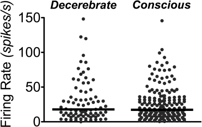
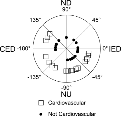
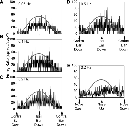
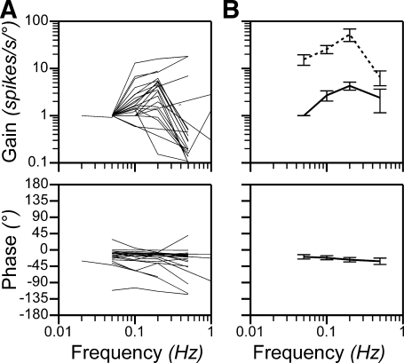
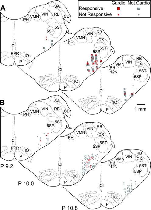
The responses to vestibular stimulation of brain stem neurons that regulate sympathetic outflow and blood flow have been studied extensively in decerebrate preparations, but not in conscious animals. In the present study, we compared the responses of neurons in the rostral ventrolateral medulla (RVLM), a principal region of the brain stem involved in the regulation of blood pressure, to whole body rotations of conscious and decerebrate cats. In both preparations, RVLM neurons exhibited similar levels of spontaneous activity (median of ∼17 spikes/s). The firing of about half of the RVLM neurons recorded in decerebrate cats was modulated by rotations; these cells were activated by vertical tilts in a variety of directions, with response characteristics suggesting that their labyrinthine inputs originated in otolith organs. The activity of over one-third of RVLM neurons in decerebrate animals was altered by stimulation of baroreceptors; RVLM units with and without baroreceptor signals had similar responses to rotations. In contrast, only 6% of RVLM neurons studied in conscious cats exhibited cardiac-related activity, and the firing of just 1% of the cells was modulated by rotations. These data suggest that the brain stem circuitry mediating vestibulosympathetic reflexes is highly sensitive to changes in body position in space but that the responses to vestibular stimuli of neurons in the pathway are suppressed by higher brain centers in conscious animals. The findings also raise the possibility that autonomic responses to a variety of inputs, including those from the inner ear, could be gated according to behavioral context and attenuated when they are not necessary.
Figures
Similar articles
-
Responses of neurons in the rostral ventrolateral medulla of the cat to natural vestibular stimulation.Brain Res. 1993 Jan 22;601(1-2):255-64. doi: 10.1016/0006-8993(93)91718-8. Brain Res. 1993. PMID: 8431771
-
Responses of neurons in the rostral ventrolateral medulla of conscious cats to anticipated and passive movements.Am J Physiol Regul Integr Comp Physiol. 2020 Mar 1;318(3):R481-R492. doi: 10.1152/ajpregu.00205.2019. Epub 2020 Jan 15. Am J Physiol Regul Integr Comp Physiol. 2020. PMID: 31940234 Free PMC article.
-
Processing of vestibular inputs by the medullary lateral tegmental field of conscious cats: implications for generation of motion sickness.Exp Brain Res. 2013 Mar;225(3):349-59. doi: 10.1007/s00221-012-3376-1. Epub 2012 Dec 29. Exp Brain Res. 2013. PMID: 23274644 Free PMC article.
-
Excitatory inputs to the RVLM in the context of the baroreceptor reflex.Ann N Y Acad Sci. 2001 Jun;940:247-58. doi: 10.1111/j.1749-6632.2001.tb03681.x. Ann N Y Acad Sci. 2001. PMID: 11458682 Review.
-
The baroreflex and beyond: control of sympathetic vasomotor tone by GABAergic neurons in the ventrolateral medulla.Clin Exp Pharmacol Physiol. 2002 May-Jun;29(5-6):514-21. doi: 10.1046/j.1440-1681.2002.03665.x. Clin Exp Pharmacol Physiol. 2002. PMID: 12010201 Review.
Cited by
-
Collateralization of projections from the rostral ventrolateral medulla to the rostral and caudal thoracic spinal cord in felines.Exp Brain Res. 2012 Jul;220(2):121-33. doi: 10.1007/s00221-012-3122-8. Epub 2012 May 24. Exp Brain Res. 2012. PMID: 22623097 Free PMC article.
-
Why can't rodents vomit? A comparative behavioral, anatomical, and physiological study.PLoS One. 2013 Apr 10;8(4):e60537. doi: 10.1371/journal.pone.0060537. Print 2013. PLoS One. 2013. PMID: 23593236 Free PMC article.
-
Development of manganese-enhanced magnetic resonance imaging of the rostral ventrolateral medulla of conscious rats: Importance of normalization and comparison with other regions of interest.NMR Biomed. 2018 Mar;31(3):10.1002/nbm.3887. doi: 10.1002/nbm.3887. Epub 2018 Jan 12. NMR Biomed. 2018. PMID: 29327782 Free PMC article.
-
Responses of neurons in the caudal medullary lateral tegmental field to visceral inputs and vestibular stimulation in vertical planes.Am J Physiol Regul Integr Comp Physiol. 2012 Nov 1;303(9):R929-40. doi: 10.1152/ajpregu.00356.2012. Epub 2012 Sep 5. Am J Physiol Regul Integr Comp Physiol. 2012. PMID: 22955058 Free PMC article.
-
Integration of vestibular and gastrointestinal inputs by cerebellar fastigial nucleus neurons: multisensory influences on motion sickness.Exp Brain Res. 2014 Aug;232(8):2581-9. doi: 10.1007/s00221-014-3898-9. Epub 2014 Mar 28. Exp Brain Res. 2014. PMID: 24677139 Free PMC article.
References
-
- Anderson JH, Blanks RHI, Precht W. Response characteristics of semicircular canal and otolith systems in the cat. I. Dynamic responses of primary vestibular fibers. Exp Brain Res 32: 491–507, 1978 - PubMed
-
- Barman SM, Gebber GL. Axonal projection patterns of ventrolateral medullospinal sympathoexcitatory neurons. J Neurophysiol 53: 1551–1566, 1985 - PubMed
-
- Barman SM, Gebber GL. Sequence of activation of ventrolateral and dorsal medullary sympathetic neurons. Am J Physiol Regul Integr Comp Physiol 245: R438–R477, 1983 - PubMed
-
- Bent LR, Bolton PS, Macefield VG. Modulation of muscle sympathetic bursts by sinusoidal galvanic vestibular stimulation in human subjects. Exp Brain Res 174: 701–711, 2006 - PubMed
-
- Berman AI. The Brain Stem of the Cat. Madison: University of Wisconsin Press, 1968
Publication types
MeSH terms
Grants and funding
LinkOut - more resources
Full Text Sources
Medical
Miscellaneous