. 2011 May;22(5):975-83.
doi: 10.1681/ASN.2010070777. Epub 2011 Apr 14.

Donor-specific antibodies accelerate arteriosclerosis after kidney transplantation

Affiliations

Donor-specific antibodies accelerate arteriosclerosis after kidney transplantation

Gary S Hill et al. J Am Soc Nephrol. 2011 May.

Abstract

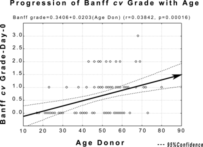
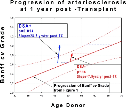
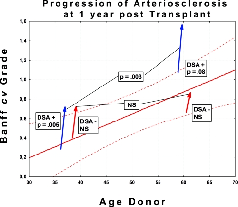
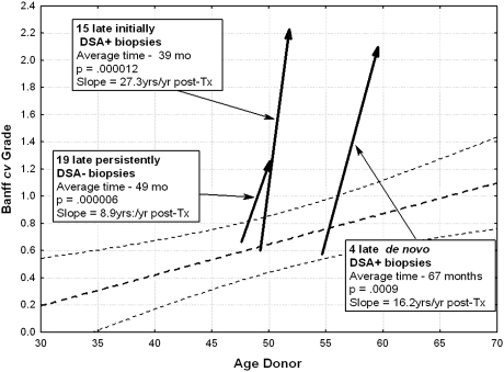
In biopsies of renal allografts, arteriosclerosis is often more severe than expected based on the age of the donor, even without a history of rejection vasculitis. To determine whether preformed donor-specific antibodies (DSAs) may contribute to the severity of arteriosclerosis, we examined protocol biopsies from patients with (n=40) or without (n=59) DSA after excluding those with any evidence of vasculitis. Among DSA-positive patients, arteriosclerosis significantly progressed between month 3 and month 12 after transplant (mean Banff cv score 0.65 ± 0.11 to 1.12 ± 0.10, P=0.014); in contrast, among DSA-negative patients, we did not detect a statistically significant progression during the same timeframe (mean Banff cv score 0.65 ± 0.11 to 0.81 ± 0.10, P=not significant). Available biopsies at later time points supported a rate of progression of arteriosclerosis in DSA-negative patients that was approximately one third that in DSA-positive patients. Accelerated arteriosclerosis was significantly associated with peritubular capillary leukocytic infiltration, glomerulitis, subclinical antibody-mediated rejection, and interstitial inflammation. In conclusion, these data support the hypothesis that donor-specific antibodies dramatically accelerate post-transplant progression of arteriosclerosis.


Figures

Figure 1.
Figure 1.
Progression of Banff cv grade at 1 year post-TX, based on 91 d-0 biopsies. The regression line here and the confidence intervals will be used in subsequent graphs showing acceleration of arteriosclerosis, using as the point of depart the mean donor age at outset.
Figure 2.
Figure 2.
Lesions progress more rapidly in DSA+ patients than in DSA− patients. Figure shows overall progression of Banff cv grade at 1 year post-TX. The 89 patients in this study were only analyzed completely for the first 12 months. The arrows begin at the median donor age for the cohort in question.
Figure 3.
Figure 3.
There are no significant differences in rate of progression between patients over 50 years of age compared with those below 50 years of age. Figure shows progression of Banff cv grade at 1 year post-TX. Cohorts are divided into those ≤50 and <50 years of age. The arrows begin at the median donor age for the cohort in question.
Figure 4.
Figure 4.
Patients with de novo positivity had rates of progression more nearly approaching that of initially DSA+ cases than that of persistently DSA− patients. Figure shows progression of Banff cv in late biopsies. The arrows begin at the median donor age for the cohort in question.
Figure 5.
Figure 5.
Morphologic features of accelerated arteriosclerosis. (A) Artery without preexisting arteriosclerosis, 3 months after TX. Several cells (arrows) interposed between internal elastica and endothelim, with little associated extracellular matrix. Such cells stain positively for α-SMA. Masson trichrome stain (MT), ×550. (B) Artery with prior arteriosclerosis, 3 months after TX. Several cells lying immediately beneath the endothelium. The amount of extracellular matrix they have elaborated is not possible to determine. MT, ×500. (C) Artery at 1 year after TX. The intima is markedly thickened and is quite cellular throughout with abundant relatively mature collagen fibers. MT, ×400. (D) Arteries at 1 year after TX. The left panel shows an artery stained for elastic fibers, showing well-formed layer adjacent to endothelium with prominent cells (arrow) just beneath in the thickened intima. Orcein stain, ×550. Right panel shows artery stained for α-SMA showing hypercellular intima with abundant positivity. Arrow shows the gray-staining internal elastica. Everything within represents expanded intima. Magnification, ×600. (E) Artery at 2 years after TX. The outer portions of the intima are composed of dense collagen, but the inner portion remains hypercellular, although collagen appears to be relatively mature. Patient had active ongoing SAMR. MS, ×350. (F) Artery at 3 years after TX. Distinction between relatively hypercellular inner intimal zone and dense collagen in outer intima remains. Patient had active SAMR. Magnification, ×350 (G) Artery at 3 years. Dense collagenous intima shows only modest cellularity. Patient currently had only minimal evidence of AMR (minimal glomerulitis graded as ± with no PTC infiltration). MT, ×350. (H) Artery in DSA− patient at 55 months. The intima is mildly hypercellular, the elongated cells lying between bands of mature collagen. MT,×350.

Similar articles

Cited by

References

    1. Colvin RB: Antibody-mediated renal allograft rejection: Diagnosis and pathogenesis. J Am Soc Nephrol 18: 1046–1056, 2007 - PubMed
    1. Colvin RB: Renal transplant pathology. In: Heptinstall's Pathology of the Kidney, 6th ed., edited by Jennette JC, Olson JL, Schwartz MM, Silva FG. Philadelphia, Lippincott, Williams & Wilkins, 2007, pp 1347–1490
    1. Cornell LD, Smith RN, Colvin RB: Kidney transplantation: Mechanisms of rejection and acceptance. Annu Rev Pathol 3: 189–220, 2008 - PubMed
    1. Racusen LC, Colvin RB, Solez K, Mihatsch MJ, Halloran PF, Campbell PM, Cecka MJ, Cosyns JP, Demetris AJ, Fishbein MC, Fogo A, Furness P, Gibson IW, Glotz D, Hayry P, Hunsickern L, Kashgarian M, Kerman R, Magil AJ, Montgomery R, Morozumi K, Nickeleit V, Randhawa P, Regele H, Seron D, Seshan S, Sund S, Trpkov K: Antibody-mediated rejection criteria: An addition to the Banff 97 classification of renal allograft rejection. Am J Transplant 3: 708–714, 2003 - PubMed
    1. Lefaucheur C, Nochy D, Hill GS, Glotz: Are transplant endarteritis (v1, v2) part of the spectrum of antibody-mediated rejection? [Abstract] 9th Banff Conference on Allograft Pathology, La Coruña, Spain, 2007