. 2011 May;22(5):902-13.
doi: 10.1681/ASN.2010070705. Epub 2011 Apr 14.

Autophagy protects the proximal tubule from degeneration and acute ischemic injury

Affiliations

Autophagy protects the proximal tubule from degeneration and acute ischemic injury

Tomonori Kimura et al. J Am Soc Nephrol. 2011 May.

Abstract

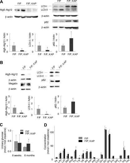
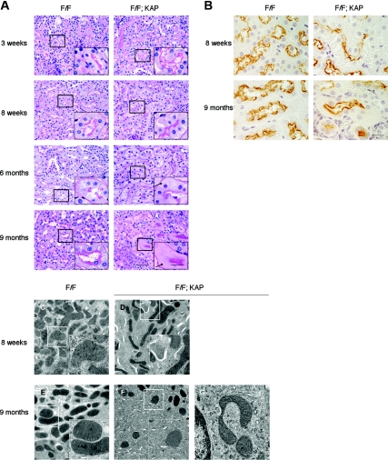
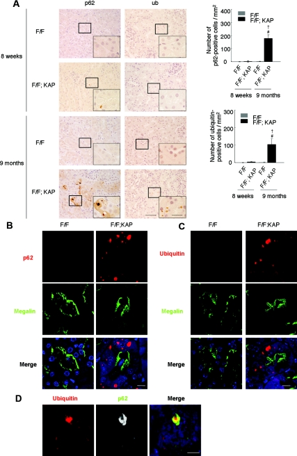
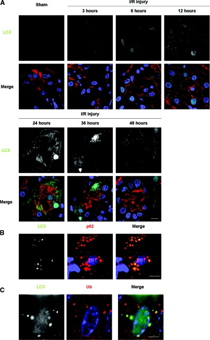
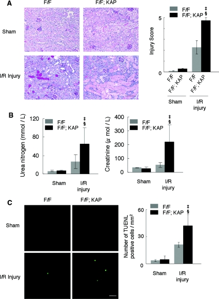
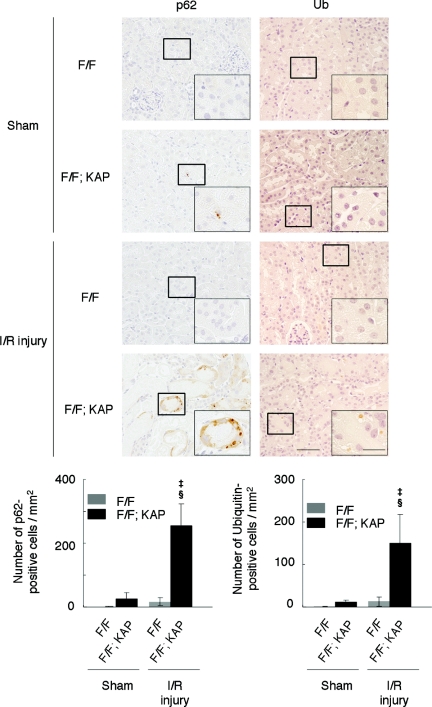
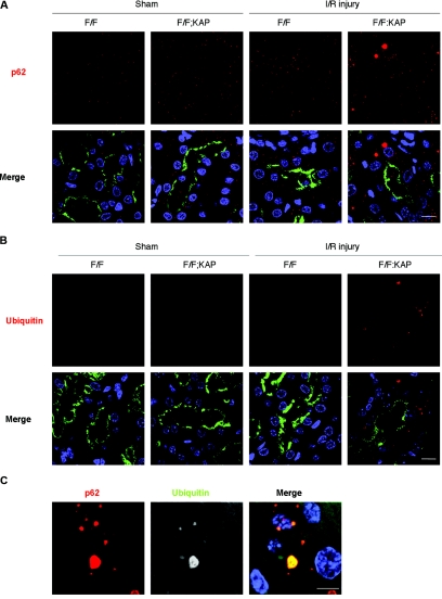
Autophagy is a bulk protein degradation system that likely plays an important role in normal proximal tubule function and recovery from acute ischemic kidney injury. Using conditional Atg5 gene deletion to eliminate autophagy in the proximal tubule, we determined whether autophagy prevents accumulation of damaged proteins and organelles with aging and ischemic renal injury. Autophagy-deficient cells accumulated deformed mitochondria and cytoplasmic inclusions, leading to cellular hypertrophy and eventual degeneration not observed in wildtype controls. In autophagy-deficient mice, I/R injury increased proximal tubule cell apoptosis with accumulation of p62 and ubiquitin positive cytoplasmic inclusions. Compared with control animals, autophagy-deficient mice exhibited significantly greater elevations in serum urea nitrogen and creatinine. These data suggest that autophagy maintains proximal tubule cell homeostasis and protects against ischemic injury. Enhancing autophagy may provide a novel therapeutic approach to minimize acute kidney injury and slow CKD progression.


Figures

Figure 1.
Figure 1.
Physiologic changes in proximal tubule-specific autophagy-deficient mice. (A and B) Western blot analysis confirmed Atg5 deficiency, suppression of the conversion of LC3-I to LC3-II, and accumulation of p62 in (A) kidney cortexes of 8-week-old Atg5flox/flox; KAP-Cre+ (F/F; KAP) mice (n = 5) and in (B) lysates of isolated proximal tubular cells of 3-week-old Atg5flox/flox; KAP-Cre+ mice (n = 3 to 4). A representative immunoblot is shown. The graph shows the ratios of band intensities of Atg5-Atg12, LC3-II, and p62 to those of actin, standardized to the mean of the control. Megalin was blotted as a marker of the proximal tubular cells. (C) Glycosiuria was observed in 6-month-old Atg5flox/flox; KAP-Cre+ mice (n = 5 to 6). (D) Mild amino aciduria was observed in 6-month-old Atg5flox/flox; KAP-Cre+ mice (n = 5 to 6). Cysteine and serine were not detected. Data are presented as means ± SD. *P < 0.05 versus Atg5flox/flox; KAP-Cre mice of same age.
Figure 2.
Figure 2.
Histologic changes in proximal tubule-specific autophagy-deficient mice. (A) PAS-stained kidney cortex from 3-week-old, 8-week-old, 6-month-old, and 9-month-oldAtg5flox/flox; KAP-Cre (left) and Atg5flox/flox; KAP-Cre+ (right) mice (n = 4 to 5). Atg5flox/flox; KAP-Cre+ mice exhibited slight hypertrophy of the tubular cells at 8 weeks and accumulation of cytosolic amorphous substrates at 6 months. Arrows indicate tubular cells with cytosolic accumulation of amorphous substrates. Bars, 50 and 20 μm (insets). (B) Immunostaining of megalin, a marker of the proximal tubular brush border, showed that the hypertrophied cells (8 weeks) and amorphous substrates-accumulated cells (9 months) are proximal tubular cells in Atg5flox/flox; KAP-Cre+ mice (n = 5). Bar, 20 μm. (C–E) Atg5 deficiency causes accumulation of crescent-like structures and deformed mitochondria. Electron micrographs of kidney proximal tubules from Atg5flox/flox; KAP-Cre (C and E) and Atg5flox/flox; KAP-Cre+ (D, F, and G) mice of 8 weeks (C and D) and 9 months (E through G) of age. The autophagy-deficient proximal tubular cells showed accumulation of crescent-like structures in 8-week-old (D) and 9-month-old (F) mice. (G) The autophagy-deficient proximal tubular cells of 9-month-old mice also contained deformed mitochondria. The figure is a representative of multiple experiments (n = 3). Bars, 2 μm (C–F) and 500 nm (insets of C–F and G). Magnification, ×400 (A), ×1000 (insets of A and B), ×2500 (C through F), and ×10,000 (insets of C–F and G).
Figure 3.
Figure 3.
Atg5 deficiency causes accumulation of p62- and ubiquitin-positive inclusions in the kidney cortex tubular cells of 9-month-old mice. (A) Immunohistologic analysis of Atg5flox/flox; KAP-Cre and Atg5flox/flox; KAP-Cre+ mice kidney cortexes from 8-week-old and 9-month-old mice. Kidney tubular cells of 8-week-old mice showed little staining of p62 (left) or ubiquitin (right), whereas those of 9-month-old mice showed massive accumulation of p62- and ubiquitin -positive inclusions in Atg5flox/flox; KAP-Cre+ mice. These inclusions were not obsereved in Atg5flox/flox; KAP-Cre mice. P < 0.05 versus 8-week-old Atg5flox/flox; KAP-Cre+ mice; #P < 0.05 versus 9-month-old Atg5flox/flox; KAP-Cre mice. Data are presented as means ± SD. (B and C) Immunofluorescence analysis showed that marked accumulation of p62-positive (B) and ubiquitin-positive (C) large dots was observed in autophagy-deficient kidney proximal tubules of 9-month-old mice. Megalin was used as a marker of the proximal tubules. DAPI staining was performed as a counterstaining. (D) p62 and ubiquitin colocalized in the inclusions of autophagy-deficient kidney proximal tubules of 9-month-old mice. The figure is representative of a multiple experiments (n = 5). Bars, 50 (A), 20 (insets of A), 10 (B and C), and 5μm (D). Magnification, ×400 (A), ×1000 (Inlets), ×352 (B and C), and ×1058 (D).
Figure 4.
Figure 4.
Formation of LC3 dots in kidney proximal tubules of LC3-GFP transgenic mice in response to I/R injury. (A) The presence of LC3-positive dots in the proximal tubules of the kidney of LC3-GFP transgenic mice was examined by immunofluorescence analysis after a sham operation and after 3, 6, 12, 24, 36, and 48 hours after unilateral I/R injury. The number of LC3 dots increased time-dependently after injury for up to 24 hours, after which it decreased. Lotus tetragonolobus lectin (LTA) was used as a marker of proximal tubules, and DAPI staining was performed as a counterstaining. (B and C) LC3 dots colocalized with p62 (B) and ubiquitin dots (C) after I/R injury. Kidney tissues 12 hours after unilateral I/R injury is shown. The figure is representative of a multiple experiments (n = 3 to 5). Bars, 10 (A) and 5 μm (B and C). Magnification, ×352 (A), ×1058 (B), and ×1408 (C).
Figure 5.
Figure 5.
Marked injury in autophagy-deficient kidney cortex after I/R injury. (A) Histologic analysis of Atg5flox/flox; KAP-Cre (left) and Atg5flox/flox; KAP-Cre+ (right) kidneys 2 days after unilateral I/R injury (n = 5). PAS-stained kidney cortexes showed that the tubules of Atg5flox/flox; KAP-Cre+ mice were severely injured compared with I/R-injured Atg5flox/flox mice. The tubular injury score of the Atg5flox/flox; KAP-Cre+ mice was significantly increased compared with that of controls. (B) Bilateral I/R injury caused significant increase of serum urea nitrogen and creatinine in Atg5flox/flox; KAP-Cre+ mice compared with I/R-injured control littermates (n = 4 to 6). (C) Terminal deoxynucleotidyl transferase-mediated deoxyuridine triphosphate nick end-labeling staining 2 days after I/R injury showed increased number of apoptotic tubular cells in the cortex of Atg5flox/flox; KAP-Cre+ mice (n = 5). P < 0.05 versus sham-operated Atg5flox/flox; KAP-Cre+ mice; §P < 0.05 versus I/R injury-operated Atg5flox/flox; KAP-Cre mice. Data are presented as means ± SD. Bars: 100 (A) and 10 μm (C). The images are representatives of multiple experiments. Magnification, ×200 (A) and ×352 (C).
Figure 6.
Figure 6.
Accumulation of p62- and ubiquitin-positive inclusions in the autophagy-deficient kidney cortex after I/R injury. Immunohistologic analysis of Atg5flox/flox; KAP-Cre and Atg5flox/flox; KAP-Cre+ kidneys 2 days after I/R injury. p62 (left) or ubiquitin (right) staining of kidney cortexes showed massive accumulation of p62- and ubiquitin-positive inclusions in Atg5flox/flox; KAP-Cre+ kidneys after I/R injury. P < 0.05 versus sham-operated Atg5flox/flox; KAP-Cre+ mice; §P < 0.05 versus I/R injury-operated Atg5flox/flox; KAP-Cre mice. Data are presented as means ± SD. The figure is a representative of multiple experiments (n = 5). Bars, 50 and 20 μm (insets). Magnification, ×400 and ×1000 (insets).
Figure 7.
Figure 7.
Formation of p62- and ubiquitin-positive inclusions in autophagy-deficient kidney proximal tubules in response to I/R injury. (A and B) Immunofluorescence analysis showed that marked accumulation of p62-positive large dots (A) and ubiquitin-positive dots (B) was observed in autophagy-deficient kidney proximal tubules 2 days after I/R injury. (C) p62 and ubiquitin colocalized in the inclusions of autophagy-deficient kidney proximal tubules 2 days after I/R injury. Megalin was stained as a marker of proximal tubules in A and B. The figure is a representative of multiple experiments (n = 5). Bars, 10 (A and B) and 5 μm (C). Magnification, ×352 (A and B) and ×1058 (C).

Similar articles

Cited by

References

    1. Mizushima N, Ohsumi Y, Yoshimori T: Autophagosome formation in mammalian cells. Cell Struct Funct 27: 421–429, 2002 - PubMed
    1. Levine B, Klionsky DJ: Development by self-digestion: molecular mechanisms and biological functions of autophagy. Dev Cell 6: 463–477, 2004 - PubMed
    1. Mizushima N, Levine B, Cuervo AM, Klionsky DJ: Autophagy fights disease through cellular self-digestion. Nature 451: 1069–1075, 2008 - PMC - PubMed
    1. Levine B, Kroemer G: Autophagy in the pathogenesis of disease. Cell 132: 27–42, 2008 - PMC - PubMed
    1. Hara T, Nakamura K, Matsui M, Yamamoto A, Nakahara Y, Suzuki-Migishima R, Yokoyama M, Mishima K, Saito I, Okano H, Mizushima N: Suppression of basal autophagy in neural cells causes neurodegenerative disease in mice. Nature 441: 885–889, 2006 - PubMed

Publication types

Substances