. 2011 Nov;45(5):977-83.
doi: 10.1165/rcmb.2011-0001OC. Epub 2011 Apr 14.

Radical-containing particles activate dendritic cells and enhance Th17 inflammation in a mouse model of asthma

Affiliations

Radical-containing particles activate dendritic cells and enhance Th17 inflammation in a mouse model of asthma

Pingli Wang et al. Am J Respir Cell Mol Biol. 2011 Nov.

Abstract

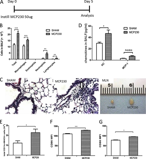
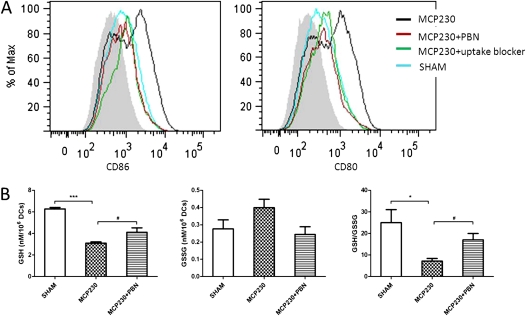
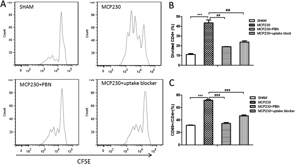
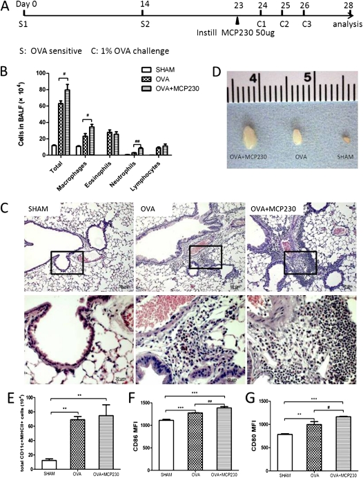
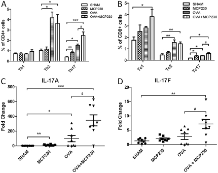
We identified a previously unrecognized component of airborne particulate matter (PM) formed in combustion and thermal processes, namely, environmentally persistent free radicals (EPFRs). The pulmonary health effects of EPFRs are currently unknown. In the present study, we used a model EPFR-containing pollutant-particle system referred to as MCP230. We evaluated the effects of MCP230 on the phenotype and function of bone marrow-derived dendritic cells (BMDCs) in vitro and lung dendritic cells (DCs) in vivo, and the subsequent T-cell response. We also investigated the adjuvant role of MCP230 on airway inflammation in a mouse model of asthma. MCP230 decreased intracellular reduced glutathione (GSH) and the GSH/oxidized glutathione ratio in BMDCs, and up-regulated the expression of costimulatory molecules CD80 and CD86 on DCs. The maturation of DCs was blocked by inhibiting oxidative stress or the uptake of MCP230. BMDCs exposed to MCP230 increased their antigen-specific T-cell proliferation in vitro. In a model of asthma, exposure to MCP230 exacerbated pulmonary inflammation, which was attributed to the increase of neutrophils and macrophages but not eosinophils. This result correlated with an increase in Th17 cells and cytokines, compared with non-MCP230-treated but ovalbumin (OVA)-challenged mice. The percentage of Th2 cells was comparable between OVA and OVA + MCP230 mice. Our data demonstrate that combustion-generated, EPFR-containing PM directly induced the maturation of DCs in an uptake-dependent and oxidative stress-dependent manner. Furthermore, EPFR-containing PM induced a Th17-biased phenotype in lung, accompanied by significant pulmonary neutrophilia. Exposure to EPFR-containing PM may constitute an important and unrecognized risk factor in the exacerbation and development of a severe asthma phenotype in humans.


Figures

Figure 1.
Figure 1.
Exposure to the environmentally persistent free radical (EPFR)–containing pollutant-particle system MCP230 induced an innate immune response in lungs and the maturation of pulmonary dendritic cells (DCs). (A) Mice received MCP230 via oropharyngeal aspiration on Day 0. Bronchoalveolar lavage fluid (BALF) and lungs were isolated for analyses of cellularity and chemokines on Day 5. (B) Exposure to MCP230 increased the total numbers of leukocytes, neutrophils, monocytes, and lymphocytes in BALF. (C) Representative micrographs demonstrate inflammatory cell infiltrates in the perivascular and peribronchiolar regions (arrow). The mediastinal lymph node (MLN) was enlarged upon instillation of MCP230. (D) Exposure to MCP230 increased concentrations of chemokine (C-X-C motif) ligand 1 (KC) and MCP–1 in BALF. (E) Exposure to MCP230 increased the total number of lung DCs (CD11c+MHCII+ cells). (F and G) The mean fluorescence intensity (MFI) of CD86 (F) and CD80 (G) in lung DCs was determined by flow cytometry. *P < 0.05. **P < 0.01. ***P < 0.001.
Figure 2.
Figure 2.
Maturation of DCs and oxidative stress after exposure to MCP230 in vitro. (A) Expression of costimulatory molecules in CD11c+ MHCII+ bone marrow–derived dendritic cells (BMDCs). BMDCs were exposed to vehicle (SHAM) or MCP230, or pretreated with PBN or uptake blocker. Twenty-four hours later, the expression of CD86 and CD80 in CD11c+ MHCII+ BMDCs was determined by flow cytometry. Gray area in histogram represents isotype control. (B) MCP230 regulates BMDC glutathione homeostasis. After exposure to MCP230 for 24 hours, DCs were collected and lysed. Intracellular reduced glutathione (GSH) and oxidized glutathione (GSSG) were measured in cell lysates. *P < 0.05. #P < 0.05. ***P < 0.001.
Figure 3.
Figure 3.
MCP230 alters the capacity of DCs to prime T cells. BMDCs were exposed to MCP230 or vehicle (SHAM) and ovalbumin peptide (OVA323–339), with or without pretreatment with PBN or uptake blocker, for 24 hours. DCs were then cocultured with carboxyfluorescein diacetate, succinimidyl ester (CFSE)-labeled naive DO11.10 TCR+CD4+ T cells for 72 hours. Fluorescence intensities were measured by flow cytometry. (A) Representative histogram of CFSE division profiles of CD4+ T cells in each group. Data indicate percentages of dividing CD4+ T cells (B) and CD69+ activated CD4+ T cells (C). ##P < 0.01. ###P < 0.001. ***P < 0.001.
Figure 4.
Figure 4.
MCP230 enhanced pulmonary inflammation in a mouse model of asthma by inducing the maturation of DCs. (A) Schematic of the protocol used to induce allergic inflammation (i.e., mouse model of asthma). OVA complexed to Imject Alum was administered intraperitoneally on Protocol Days 0 and 14. Mice were then challenged with aerosolized OVA on Protocol Days 24, 25, and 26. The OVA + MCP230 group was exposed to MCP230 on Protocol Day 23. Sham control mice were exposed to saline and vehicle. (B) BALF was acquired, and cells were differentiated and counted. (C) Representative micrographs of the lungs of mice exposed to sham, OVA, or OVA + MCP230 (hematoxylin and eosin stain). (D) MLNs from OVA + MCP230 mice were larger than those of OVA mice. (E–G) Lung cells were isolated and stained with fluorescently labeled antibodies to determine the numbers and maturation status of DCs. Numbers of pulmonary DCs (E) and the MFIs of CD86 (F) and CD80 (G) in lung DCs were determined by flow cytometry. #P < 0.05. ##P < 0.01. **P < 0.01. ***P < 0.001.
Figure 5.
Figure 5.
MCP230 influences pulmonary T-cell responses in a mouse model of asthma. (A and B) Lung cells were isolated, stimulated with phorbol myristate acetate and ionomycin, and stained with surface antibodies (CD4 and CD8) and intracellular antibodies (IFN-γ, IL-4, and IL-17A). Subsets of T cells were then quantified using flow cytometry. (C and D) The expression of IL-17A and IL-17F genes in whole-lung homogenates was tested by real-time PCR. #P < 0.05. *P < 0.05. **P < 0.01. ***P < 0.001.

Similar articles

Cited by

References

    1. Holguin F. Traffic, outdoor air pollution, and asthma. Immunol Allergy Clin North Am 2008;28:577–588 - PubMed
    1. Patel MM, Miller RL. Air pollution and childhood asthma: recent advances and future directions. Curr Opin Pediatr 2009;21:235–242 - PMC - PubMed
    1. Salam MT, Islam T, Gilliland FD. Recent evidence for adverse effects of residential proximity to traffic sources on asthma. Curr Opin Pulm Med 2008;14:3–8 - PubMed
    1. Mamessier E, Nieves A, Vervloet D, Magnan A. Diesel exhaust particles enhance T-cell activation in severe asthmatics. Allergy 2006;61:581–588 - PubMed
    1. Cass GR. Comments on sources, atmospheric levels, and characterization of airborne particulate matter. Inhal Toxicol 1995;7:765–768

Publication types

MeSH terms