. 2011 Apr 15;332(6027):354-8.
doi: 10.1126/science.1198949.

Specificity of Drosophila cytonemes for distinct signaling pathways

Affiliations

Specificity of Drosophila cytonemes for distinct signaling pathways

Sougata Roy et al. Science. .

Abstract

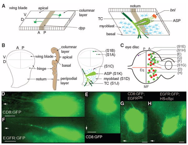
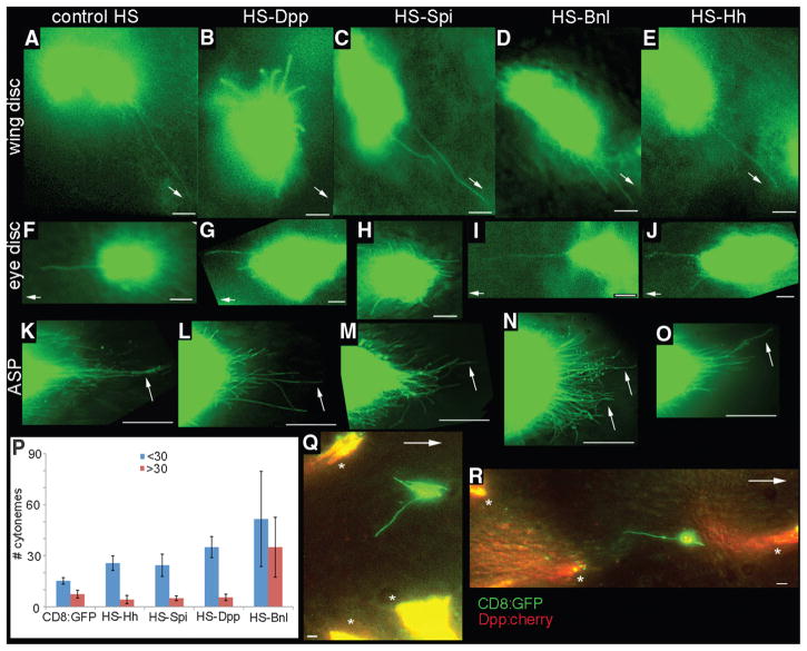
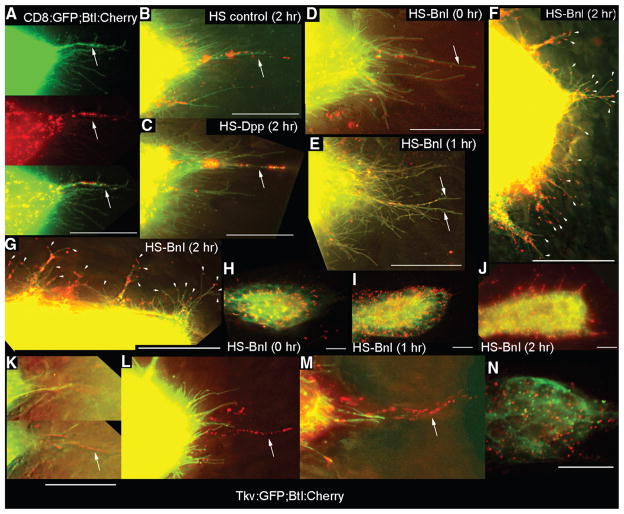
Cytonemes are types of filopodia in the Drosophila wing imaginal disc that are proposed to serve as conduits in which morphogen signaling proteins move between producing and target cells. We investigated the specificity of cytonemes that are made by target cells. Cells in wing discs made cytonemes that responded specifically to Decapentaplegic (Dpp) and cells in eye discs made cytonemes that responded specifically to Spitz (the Drosophila epidermal growth factor protein). Tracheal cells had at least two types: one made in response to Branchless (a Drosophila fibroblast growth factor protein, Bnl), to which they segregate the Bnl receptor, and another to which they segregate the Dpp receptor. We conclude that cells can make several types of cytonemes, each of which responds specifically to a signaling pathway by means of the selective presence of a particular signaling protein receptor that has been localized to that cytoneme.


Figures

Fig. 1
Fig. 1
Oriented cytonemes in the wing and eye discs. (A) Representation of cytonemes on the apical surface of the columnar layer of the wing disc (left) and of cytonemes at the basal surface of the wing disc emanating from the ASP (right). bnl− and dpp-expressing regions are in red and brown, respectively; the disc-associated Tr2 TC is partly external to the disc basal lamina (dark green) and partly within the disc basal lamina [light green (12)]; ASP is shown in light green; myoblasts, blue. (B) Drawings of third instar wing discs viewed from the columnar epithelial side (left), viewed through to the basal side (right), and viewed in cross section (middle). Positions of CD8:GFP-expressing clones cross-referenced (in parentheses) to supporting online material (SOM) figure panels. (C) Drawing of third instar eye–antennal disc with eye primordium to left, antenna primordium to right; equator, Eq; area posterior to MF, P; area anterior to MF, A. Approximate locations of CD8:GFP-expressing clones are shown [cross-referenced in parentheses to (D) to (H) and SOM figure panels] in the columnar layer with cytonemes directed toward the morphogenetic furrow (green), toward the equator (red), or in the peripodial layer (purple). Clones in overexpression genetic backgrounds are shown with small circles. (D and E) Cytonemes oriented toward the MF (D) or toward the equator (E) extended from anterior clones in the columnar layer. (F) An EGFR:GFP-expressing clone in the columnar layer illuminated puncta in cytonemes oriented to the MF. (G) A clone expressing CD8:GFP and EGFRDN extended short cytonemes. (H) EGFR:GFP-containing cytonemes radiate in many directions after ubiquitous expression of cSpi. In (D), and (F) to (H), MF is to left as indicated by arrows, and equator is down, as indicated by arrow in (E). Scale bars, 5 μm.
Fig. 2
Fig. 2
Ligand-specificity of cytonemes. Dpp, Spi, Bnl, and Hh were expressed ubiquitously by heat shock 1/2 to 2 hours before imaging CD8:GFP in columnar cell clones in the wing disc (B to E), in columnar cell clones in the eye disc (G to J), or 3 to 5 hours before imaging CD8:GFP in ASPs in which expression was driven by btl-Gal4 (K to O). CD8:GFP larvae heat-shocked although lacking the respective heat shock (HS) expression transgenes served as controls [(A), (F), and (K)]. (P) Chart tabulating number of long (>30 μm, red) and short (<30 μm, blue) cytonemes at the ASP tip (100-μm circumferential arc at the tip, average of five specimens, error bars represent SD). (Q and R) Images from wing discs with independent clones that express Dpp:Cherry (*) or CD8:GFP (green). Arrows point toward A/P signaling center [(A) to (E), (Q), and (R)], toward the MF [(F) to (J )], or to long cytonemes at the ASP tip [(K) to (O)]. Scale bars for (A) to (J ), (Q), and (R), 5 μm; for (K) to (O), 30 μm.
Fig. 3
Fig. 3
Dpp and FGF receptors segregate to separate locations in the ASP. (A to J) Expression of Btl:Cherry and CD8:GFP driven by btl-Gal4 mark ASP cytonemes (green) with Btl-containing puncta (red). [(A) and (B)] ASP tips from larvae lacking a HS transgene with (B) or without (A) heat shock. (C) Btl:Cherry puncta in ASP tip cytonemes after ectopic expression of Dpp. [(D) to (F) and (H) to (J)] Expression of Btl:Cherry and CD8:GFP released from Gal80ts repression by temperature elevation (3) and subjected to heat shock for the indicated times. (E) Image of ASP tip 1 hour after heat shock. (F) Image of ASP tip 2 hours after heat shock [also see table S4 and (3)]. (G) Image of one side of the ASP tube between the TC and tip of (F). Arrowheads in (F) and (G) indicate short cytonemes containing Btl:Cherry puncta. [(H) to (J)] Low-resolution (40×) images showing distribution of Btl:Cherry after overexpression of Bnl. (F), (G), and (J) are images of the same preparation. (K to N) Tip of ASPs expressing Tkv:GFP and Btl:Cherry driven by btl-Gal4. (K) Two focal planes from z-sections in which Tkv:GFP-containing cytonemes were in a focal plane (top) less proximal to the disc than those with Btl:Cherry (bottom). [(L) and (M)] Projection images show distinct Tkv:GFP and Btl:Cherry–containing cytonemes at ASP tip. (N) Image of mid-distal ASP. Arrows in (A) to (D) and (K) to (M) indicate long cytonemes containing Btl:Cherry puncta. All images oriented ASP tip to right. Scale bars, 30 μm.

Comment in

References

    1. Kornberg TB, Guha A. Curr Opin Genet Dev. 2007;17:264. - PMC - PubMed
    1. Ramírez-Weber FA, Kornberg TB. Cell. 1999;97:599. - PubMed
    1. Materials and methods are available as supporting material on Science Online.

    1. Yang TT, Cheng L, Kain SR. Nucleic Acids Res. 1996;24:4592. - PMC - PubMed
    1. Hsiung F, Ramirez-Weber FA, Iwaki DD, Kornberg TB. Nature. 2005;437:560. - PubMed

Publication types

MeSH terms