Review
. 2011 Aug;32(4):453-71.
doi: 10.1210/er.2010-0011. Epub 2011 Apr 14.

Pituitary stem cell update and potential implications for treating hypopituitarism

Affiliations
Review

Pituitary stem cell update and potential implications for treating hypopituitarism

Frederic Castinetti et al. Endocr Rev. 2011 Aug.

Abstract

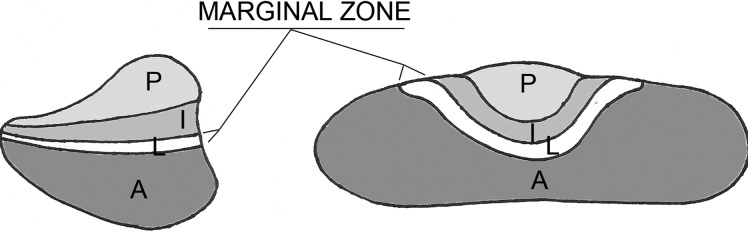
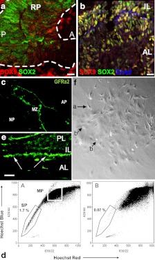
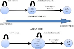
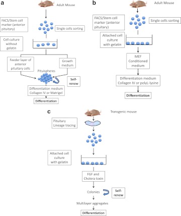
Stem cells have been identified in organs with both low and high cell turnover rates. They are characterized by the expression of key marker genes for undifferentiated cells, the ability to self-renew, and the ability to regenerate tissue after cell loss. Several recent reports present evidence for the presence of pituitary stem cells. Here we offer a critical review of the field and suggest additional studies that could resolve points of debate. Recent reports have relied on different markers, including SOX2, nestin, GFRa2, and SCA1, to identify pituitary stem cells and progenitors. Future studies will be needed to resolve the relationships between cells expressing these markers. Members of the Sox family of transcription factors are likely involved in the earliest steps of pituitary stem cell proliferation and the earliest transitions to differentiation. The transcription factor PROP1 and the NOTCH signaling pathway may regulate the transition to differentiation. Identification of the stem cell niche is an important step in understanding organ development. The niche may be the marginal zone around the lumen of Rathke's pouch, between the anterior and intermediate lobes of mouse pituitary, because cells in this region apparently give birth to all six pituitary hormone cell lineages. Stem cells have been shown to play a role in recurrent malignancies in some tissues, and their role in pituitary hyperplasia, pituitary adenomas, and tumors is an important area for future investigation. From a therapeutic viewpoint, the ability to cultivate and grow stem cells in a pituitary predifferentiation state might also be helpful for the long-term treatment of pituitary deficiencies.


Figures

Fig. 1.
Fig. 1.
Schematic representation of the marginal zone, a niche for presumed pituitary stem cells. P, Posterior lobe; I, intermediate lobe; A, anterior lobe; L, lumen.
Fig. 2.
Fig. 2.
Markers of putative pituitary stem cells. a and b, SOX9 is expressed in the embryonic pituitary gland and defines two populations of SOX2+ cells. At e12.5 (a), the majority of SOX2+ cells (green) are SOX9 negative (red) (RP, Rathke's pouch; A, anterior; P, posterior) (14). At e18.5 (b) and in adults, the majority of SOX2+ cells are SOX9+ cells (yellow staining); these cells are probably transit-amplifying cells (14). Some cells are still SOX2+, SOX9 (green) (AL, Anterior lobe; IL, intermediate lobe). E-cadherin (blue) is a marker of marginal cells and folliculostellate cells (14). c, The marginal zone of human adult pituitary contains a niche of progenitors expressing GFRa2 (15) (AP, Anterior pituitary; MZ, marginal zone; NP, neuropituitary). d, The adult mouse anterior pituitary contains a side population divided into SCA1high- and non-SCA1high - expressing cells. The SCA1high group are presumably endothelial progenitors, and non-SCA1high are presumably pituitary stem cells. Dual wave length FACS analysis reveals the presence of typical side population cells (1.7% of total living cells) in the adult anterior pituitary that are Hoechst low (d, A) and can be blocked by verapamil (d, B) (3) (SP, Side population; MP, main population). e, Nestin-GFP cells in mouse pituitary at birth. The cells are located almost exclusively in the perilumenal area of the pituitary (16) (AL, Anterior lobe; IL, intermediate lobe, PL, posterior lobe). f, Colony-forming pituitary cells in low-density culture colonies at d 8. Phase contrast low-power view showing stellate-shaped cells with long cytoplasmic processes (arrow a) and round refractile cells (arrow b). These cells express S100β and GFAP, which are folliculostellate markers (17). Panels a and b are reproduced from reference 14, with permission. © 2008, National Academy of Sciences. Panel c is reproduced from reference 15. Panel d is reproduced from reference 3, with permission. © 2005, The Endocrine Society. Panel e is reproduced from reference 16, with permission. © 2008, The National Academy of Sciences. Panel f is reproduced from reference 17, with permission. © 2005, Elsevier.
Fig. 3.
Fig. 3.
Stem cells during embryogenesis and adulthood. Pituitary stem cells differentiate during embryogenesis and form the pituitary lineages (14). These cells evolve from SOX2 to SOX9 positivity. No other marker has been evaluated during embryogenesis. Nestin does not seem to play a role at this time point because nestin+ cells begin to proliferate after birth (16). In adults, rare cells express only SOX2. The majority of adult “progenitors” are SOX2+, SOX9+ (14). It is likely that the same cells express GFRa2 (15), AMCA, and angiotensin-converting enzyme (ACE) (55) and have a little or no SCA1 expression. These cells are probably transit-amplifying cells and are capable of limited self-renewal. Nestin may be expressed in a subset of these cells, although it is not a specific marker of pituitary cells (16). Final differentiation requires spatiotemporal regulation by several transcription factors and signaling pathways (review in Ref. 40). Diff. cell, Differentiated cell.
Fig. 4.
Fig. 4.
Different ways to isolate and differentiate pituitary stem cells. a, Cells of the anterior lobe are dissociated into single cells and sorted by FACS based on specific characteristics. Side population cells display verapamil-sensitive Hoechst dye efflux capacity and/or a stem cell marker such as SCA1 and GFRa2. Cells can be seeded in dishes with medium enriched in growth factors (DMEM/Ham's F12 supplemented with growth factors) (–15). Adult anterior pituitary cells can also be used as a feeder layer (13). After 6–8 d, pituispheres appear (16). These can be handpicked individually, dispersed into single cells, and cultured to evaluate their self-renewal capacity (generation of secondary spheres). To induce differentiation, growth medium can be replaced by a specific differentiation medium. Cells are coated on growth-factor reduced Matrigel (13, 14). 4b, Anterior pituitary cells are dissociated into single cells and sorted by FACS based on a stem cell marker (AMCA) (15). Cells can be cultured on gelatin with mouse embryonic fibroblasts (MEF). To induce differentiation, growth medium can be replaced by a specific differentiation medium. Cells are coated on Collagen IV or poly-L-lysine culture slides. c, Lineage tracing involves isolation of nestin-expressing cells based on a GFP transgene (16). Cells are cultured on gelatin. After 6- to 8-d culture in fibroblast growth factor 2 (FGF2) medium and cholera toxin, colonies can be visualized. They can be handpicked individually, dispersed into single cells, and cultured to evaluate their self-renewal capacity. After multilayer aggregates formation, differentiated cells can be observed.

Similar articles

Cited by

References

    1. Murry CE, Keller G. 2008. Differentiation of embryonic stem cells to clinically relevant populations: lessons from embryonic development. Cell 132:661–680 - PubMed
    1. Yoshimura F, Harumiya K, Ishikawa H, Otsuka Y. 1969. Differentiation of isolated chromophobes into acidophils or basophils when transplanted into the hypophysiotrophic area of hypothalamus. Endocrinol Jpn 16:531–540 - PubMed
    1. Chen J, Hersmus N, Van Duppen V, Caesens P, Denef C, Vankelecom H. 2005. The adult pituitary contains a cell population displaying stem/progenitor cell and early embryonic characteristics. Endocrinology 146:3985–3998 - PubMed
    1. Vidal S, Horvath E, Kovacs K, Cohen SM, Lloyd RV, Scheithauer BW. 2000. Transdifferentiation of somatotrophs to thyrotrophs in the pituitary of patients with protracted primary hypothyroidism. Virchows Arch 436:43–51 - PubMed
    1. Thodou E, Asa SL, Kontogeorgos G, Kovacs K, Horvath E, Ezzat S. 1995. Clinical case seminar: lymphocytic hypophysitis: clinicopathological findings. J Clin Endocrinol Metab 80:2302–2311 - PubMed

Publication types