Review
. 2011 May;40(4):199-212.
doi: 10.1259/dmfr/19478385.

Dental radiographic indicators, a key to age estimation

Affiliations
Review

Dental radiographic indicators, a key to age estimation

A S Panchbhai. Dentomaxillofac Radiol. 2011 May.

Abstract

Objective: The present review article is aimed at describing the radiological methods utilized for human age identification.

Methods: The application and importance of radiological methods in human age assessment was discussed through the literature survey.

Results: Following a literature search, 46 articles were included in the study and the relevant information is depicted in the article. Dental tissue is often preserved indefinitely after death. Implementation of radiography is based on the assessment of the extent of calcification of teeth and in turn the degree of formation of crown and root structures, along with the sequence and the stages of eruption. Several radiological techniques can be used to assist in both individual and general identification, including determination of gender, ethnic group and age. The radiographic method is a simpler and cheaper method of age identification compared with histological and biochemical methods. Radiographic and tomographic images have become an essential aid for human identification in forensic dentistry, particularly with the refinement of techniques and the incorporation of information technology resources.

Conclusion: Based on an appropriate knowledge of the available methods, forensic dentists can choose the most appropriate since the validity of age estimation crucially depends on the method used and its proper application. The multifactorial approach will lead to optimum age assessment. The legal requirements also have to be considered.


Figures

Figure 1
Figure 1
Radiograph of upper and lower jaws of a fetus at the sixteenth week of intrauterine life showing the initial mineralization of deciduous incisors
Figure 2
Figure 2
Diagrammatic representation of a radiograph of a mandible of a fetus at the twenty-sixth week of intrauterine life showing advanced mineralization in anterior teeth, outline for two cusps of deciduous first molar, one cusp for deciduous second molar and the crypt of permanent first molar
Figure 3
Figure 3
Diagrammatic representation of a radiograph of a mandible of a fetus at the thirtieth week of intrauterine life showing formula image 
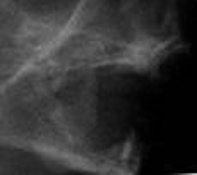
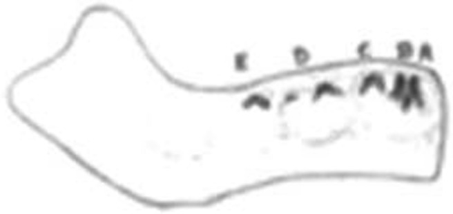
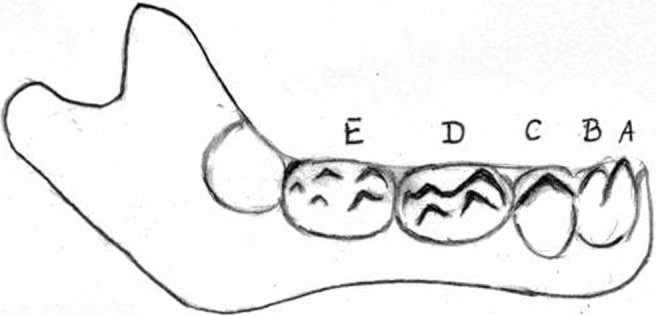
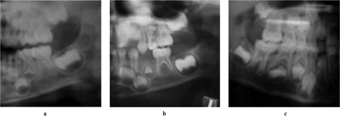
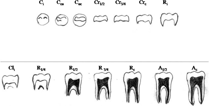
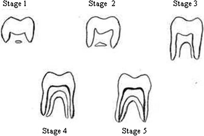
crown completion for anterior teeth, the fused cusps of deciduous first molar, five cusps of the deciduous second molar and the crypt of permanent first molar with no evidence of mineralization
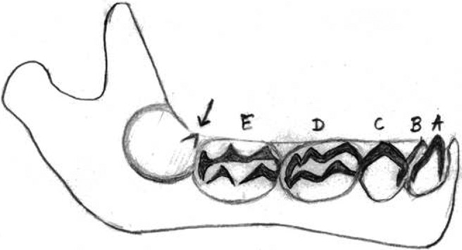
Figure 4
Figure 4
Diagrammatic representation of a radiograph of the mandible of a newborn fetus showing the completely fused cusps for deciduous first and second molar, and within the crypt of permanent first molar there is evidence of one mesial cusp tip
Figure 5
Figure 5
Developmental stages of lower deciduous first molar by Kraus and Jordan (available data). The development is described in ten stages denoted by Roman numerals from I to X; the IXth stage includes three stages and the Xth stage includes five stages
Figure 6
Figure 6
Radiographs showing upper and lower jaws with developing dentition of children aged (a) 2 years 6 months, (b) 3 years 9 months and (c) 6 years 8 months
Figure 7
Figure 7
Dental development chart by Schour and Masseler (American Dental Association, 1982), (y = years; miu = months in intrauterine life; m = months)
Figure 8
Figure 8
Cropped radiographic images showing dentition in various stages of development corresponding to given age of person (y = years, m = months)
Figure 9
Figure 9
14 stages of tooth formation of multi-rooted tooth (Moorees et al). Initial cusp formation (Ci), coalescence of cusps (Cco), cusp outline complete (Coc), crown half complete (Cr1/2), crown three quarter complete (Cr3/4), crown complete (Crc), initial root formation (Ri), initial cleft formation (Cli), root length quarter (R1/4), root length half (R1/2), root length three-quarters (R3/4), root length complete (Rc), apex half closed (A1/2), apical closure complete (Ac)
Figure 10
Figure 10
Eight stages (A to H) of mineralization of tooth (Demirjian system). A, beginning of calcification in the form of an inverted cone or cones; B, mineralized cusps are united to show coronal morphology; C, crown half formed, pulp chamber is evident; D, crown formation is completed up to cemento-enamel junction, with beginning of root formation; E, initial formation of the root bifurcation; F, the apex ends in a funnel shape; G, the walls of root canal are now parallel, its apical end partially open; H, the apical end of the root canal is completely closed; the periodontal membrane of uniform width around the root and the apex
Figure 11
Figure 11
Ten stages of tooth formation with description (Nolla's method)
Figure 12
Figure 12
Showing the measurements of tooth, L = length of tooth (L1, L2), A = distance between inner sides of open apex (A1, A2)
Figure 13
Figure 13
Diagram of premolar showing measurement sites: pulp-root length (R), pulp-tooth length (P), tooth-root length (T), pulp-root width at cemento-enamel junction (A), pulp-root width at mid-root level (C) and pulp-root width at midpoint between level C and A (B) (Kvaal et al)
Figure 14
Figure 14
Coronal length (CL) and coronal pulp cavity height or length (CPCH) for premolar and molar teeth
Figure 15
Figure 15
Five stages of lower third molar root development (Harris and Nortjè method)

Similar articles

Cited by

References

    1. Saunders E. ‘The Teeth A Test of Age’ considered with the reference to the factory children, addressed to the members of both Houses of Parliament. London: Renshaw, 1837
    1. Eckert WG, Garland N. The history of the forensic applications in radiology. Am J Forensic Med Pathol 1984;5:53–56 - PubMed
    1. Matsikidis G, Schultz P. Alterbestimmung nach dem Gebbis mit Hilfe des Zahnfilms. Zahnarztl Mitt 1982;72:2524–2528 - PubMed
    1. Ciapparelli L. The chronology of dental development and age assessment. In: Clark DH. Practical forensic odontology. Oxford: Wright Butterworth-Heinemann Ltd, 1992, pp 22–42
    1. Masthan KMK. Age and sex. Textbook of forensic odontology. New Delhi: Jaypee Brothers Medical Publishers (P) Ltd, 2009, pp 59–65