

Oral tuberculosis: unusual radiographic findings
- PMID: 21493882
- PMCID: PMC3520316
- DOI: 10.1259/dmfr/75047143
Oral tuberculosis: unusual radiographic findings
Abstract
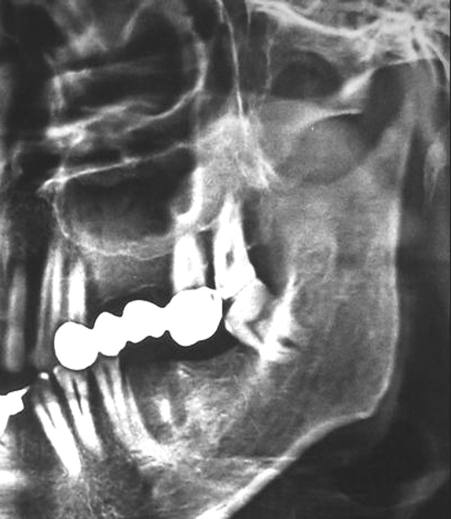
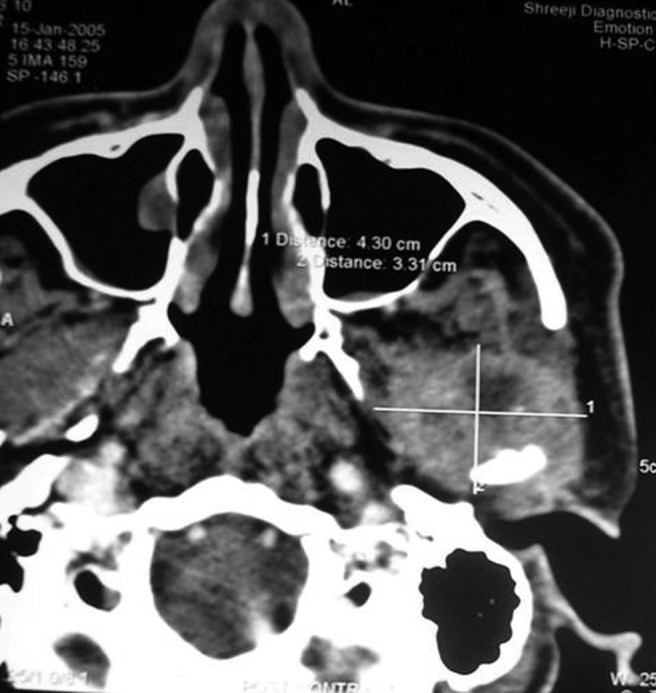
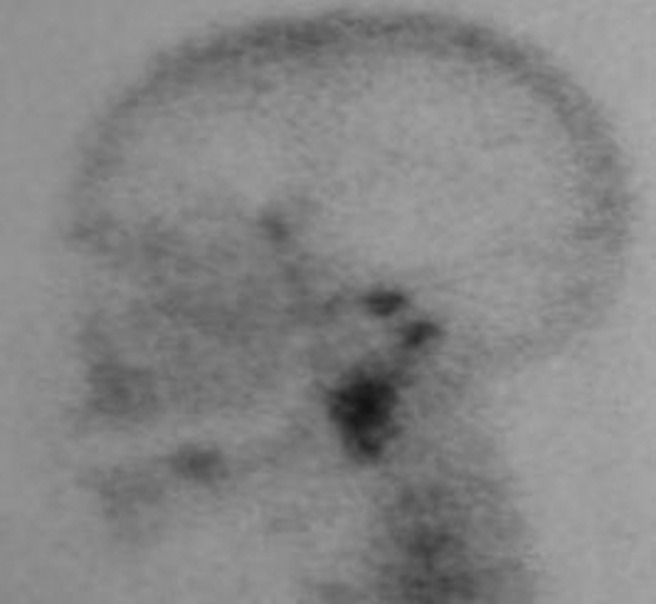
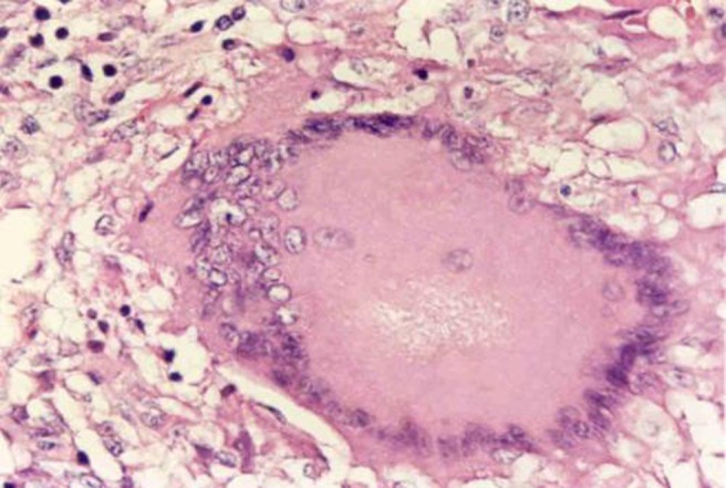
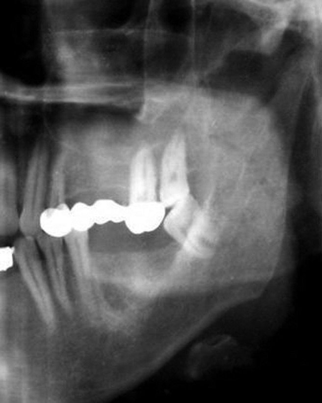
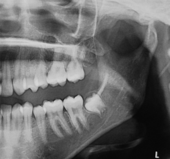
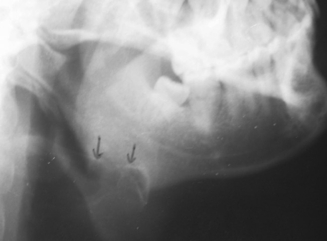
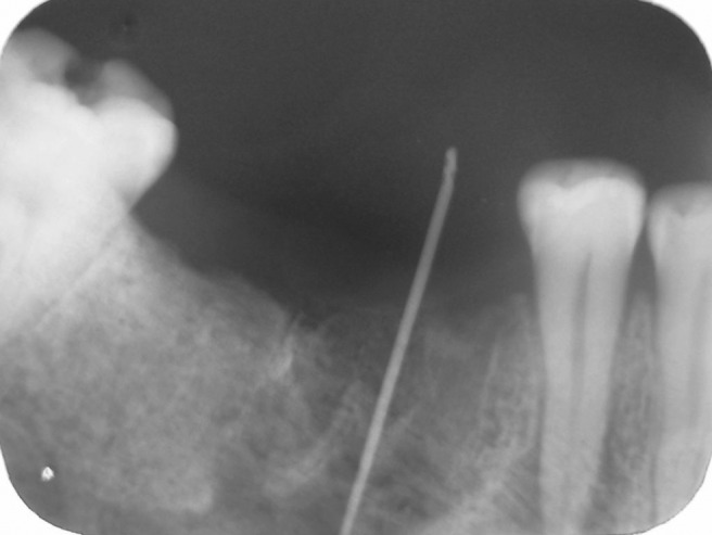
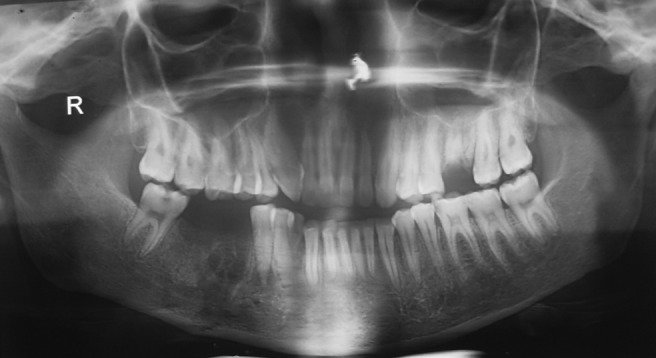
Oral tuberculosis and its radiographic findings are not commonly encountered in an oral and maxillofacial radiology practice. Literature has occasional mention of the radiographic findings of oral tuberculosis, which are still ambiguous. When affected, it is manifested majorly in the oral mucosa and rarely in the jaw bones. Here, we report certain unusual radiographic findings of oral tuberculosis which have been rarely mentioned in the literature. Four illustrative cases describe bony resorption, condylar resorption, resorption of the inferior border of the mandible and rarefaction of the alveolar bone as radiographic findings of oral tuberculosis. Follow up of the first case demonstrated regeneration of the condylar head after anti-Kochs therapy was completed, a hitherto unreported phenomenon. The importance of including tuberculosis in the differential diagnosis of some of the unusual radiographic manifestations is emphasized.
Figures
Similar articles
-
Re: Hodder et al. SPECT bone scintigraphy in the diagnosis and management of mandibular condylar hyperplasia. Br J Oral Maxillofac Surg 2000; 38: 87-93.Br J Oral Maxillofac Surg. 2001 Jun;39(3):244-5. doi: 10.1054/bjom.2000.0528. Br J Oral Maxillofac Surg. 2001. PMID: 11384126 No abstract available.
-
Condylar resorption following distraction osteogenesis: a case report.J Oral Maxillofac Surg. 2001 Sep;59(9):1104-7; discussion 1107-8. doi: 10.1053/joms.2001.25860. J Oral Maxillofac Surg. 2001. PMID: 11526589 No abstract available.
-
A comparison of single-photon emission computed tomography and planar imaging for quantitative skeletal scintigraphy of the mandibular condyle.Oral Surg Oral Med Oral Pathol Oral Radiol Endod. 1995 Aug;80(2):226-31. doi: 10.1016/s1079-2104(05)80206-9. Oral Surg Oral Med Oral Pathol Oral Radiol Endod. 1995. PMID: 7552889
-
Mandibular resorption due to systemic sclerosis. Case report of surgical correction of a secondary open bite deformity.Int J Oral Maxillofac Surg. 1995 Aug;24(4):261-7. doi: 10.1016/s0901-5027(95)80025-5. Int J Oral Maxillofac Surg. 1995. PMID: 7490486 Review.
-
Osteomyelitis of the Mandibular Condyle: A Report of 2 Cases With Review of the Literature.J Oral Maxillofac Surg. 2017 Feb;75(2):322-335. doi: 10.1016/j.joms.2016.08.018. Epub 2016 Aug 25. J Oral Maxillofac Surg. 2017. PMID: 27649464 Review.
Cited by
-
Posterior Mandibular Displacement-A Systematic Review Based on Animal Studies.Animals (Basel). 2021 Mar 15;11(3):823. doi: 10.3390/ani11030823. Animals (Basel). 2021. PMID: 33804016 Free PMC article. Review.
-
Primary labial tuberculosis: a rare presentation.Ann Med Health Sci Res. 2014 Jan;4(1):129-31. doi: 10.4103/2141-9248.126623. Ann Med Health Sci Res. 2014. PMID: 24669346 Free PMC article.
-
Indian patients' attitudes towards chairside screening in a dental setting for medical conditions.Int Dent J. 2015 Oct;65(5):269-76. doi: 10.1111/idj.12175. Epub 2015 Jul 14. Int Dent J. 2015. PMID: 26173795 Free PMC article.
-
Orofacial tuberculosis: Clinical manifestations, diagnosis and management.J Family Med Prim Care. 2015 Jul-Sep;4(3):335-41. doi: 10.4103/2249-4863.161312. J Family Med Prim Care. 2015. PMID: 26288770 Free PMC article.
-
Tubercular osteomyelitis of mandible: Defining diagnostic criteria.Natl J Maxillofac Surg. 2025 Jan-Apr;16(1):180-185. doi: 10.4103/njms.njms_56_23. Epub 2025 Apr 28. Natl J Maxillofac Surg. 2025. PMID: 40510719 Free PMC article.
References
-
- Mignogna MD, Muzio LLO, Favia G, Ruoppo E, Smmartino G, Zarrelli C, et al. Oral tuberculosis: A clinical evaluation of 42 cases. Oral Dis 2000;6:25–30 - PubMed
-
- Crompton GK, Haslett C, Chilvers ER. Diseases of the respiratory system. In: Haslett Chilvers ER, Hunter JAA, Boon NA. Davidson's principles and practice of medicine (18th edn) London: Churchill Livingstone, 1999, pp 347–353
-
- Porter R. The greatest benefit to mankind: a medical history of humanity. New York: WW Norton and Co., 1997
-
- Dye C, Scheele S, Dolin P, Pathania V, Raviglione MC. Global burden of tuberculosis: estimated incidence, prevalence, and mortality by country. J Am Med Assoc 1999;282:677–686 - PubMed
