. 2011 Jun 10;108(12):1429-38.
doi: 10.1161/CIRCRESAHA.110.237644. Epub 2011 Apr 14.

Small heat shock protein 20 interacts with protein phosphatase-1 and enhances sarcoplasmic reticulum calcium cycling

Affiliations

Small heat shock protein 20 interacts with protein phosphatase-1 and enhances sarcoplasmic reticulum calcium cycling

Jiang Qian et al. Circ Res. .

Abstract

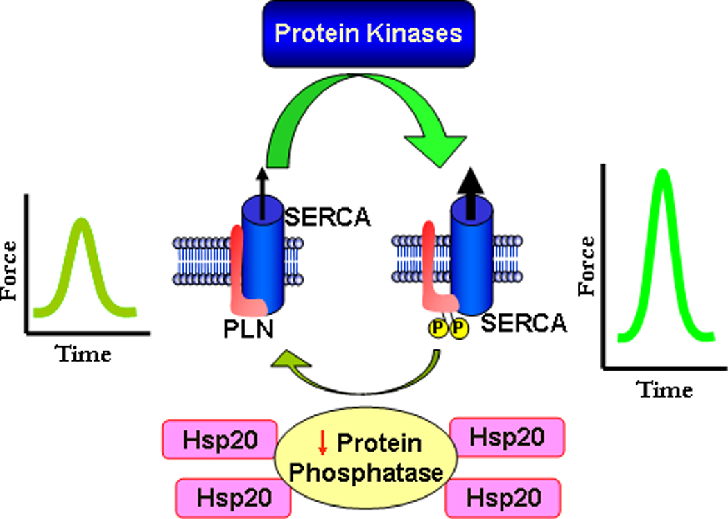
Background: Heat shock proteins (Hsp) are known to enhance cell survival under various stress conditions. In the heart, the small Hsp20 has emerged as a key mediator of protection against apoptosis, remodeling, and ischemia/reperfusion injury. Moreover, Hsp20 has been implicated in modulation of cardiac contractility ex vivo. The objective of this study was to determine the in vivo role of Hsp20 in the heart and the mechanisms underlying its regulatory effects in calcium (Ca) cycling.

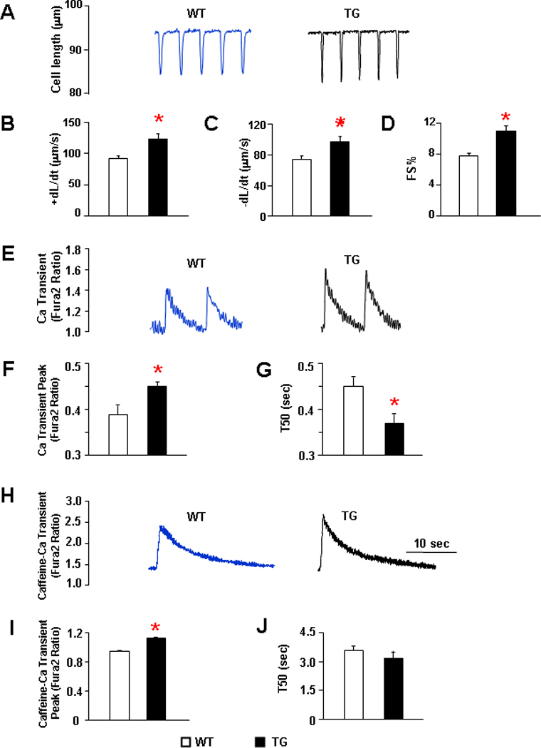
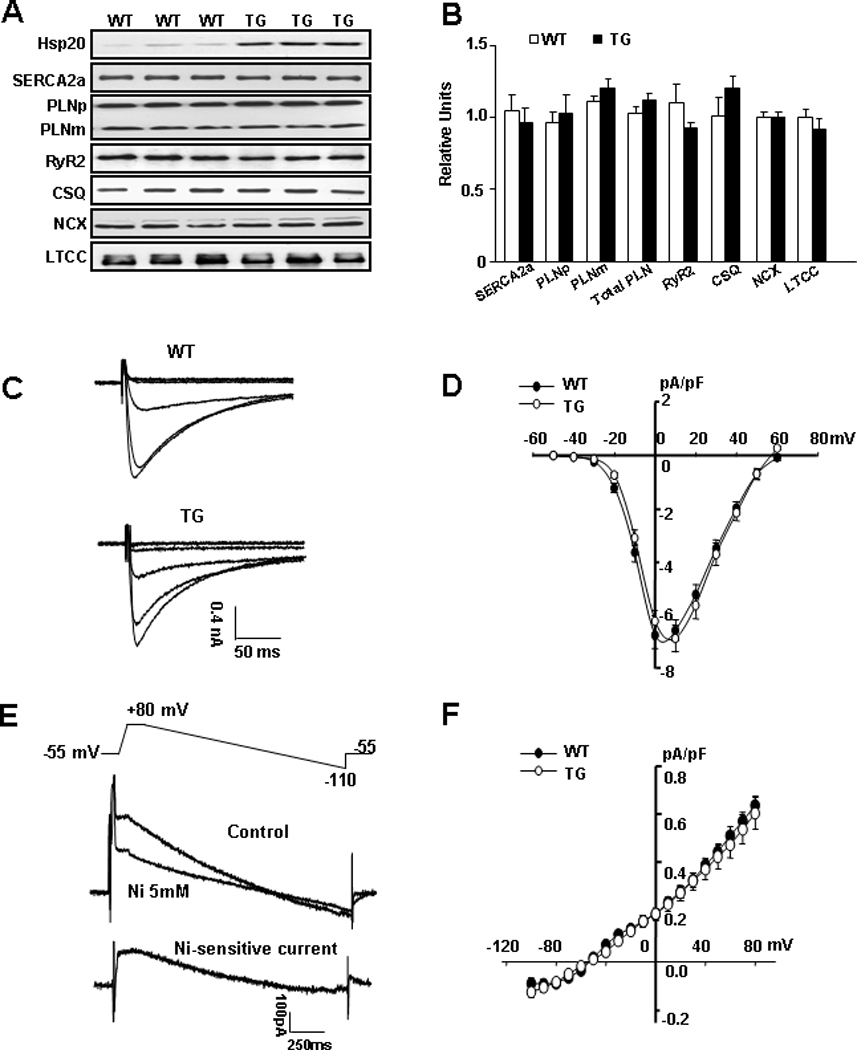
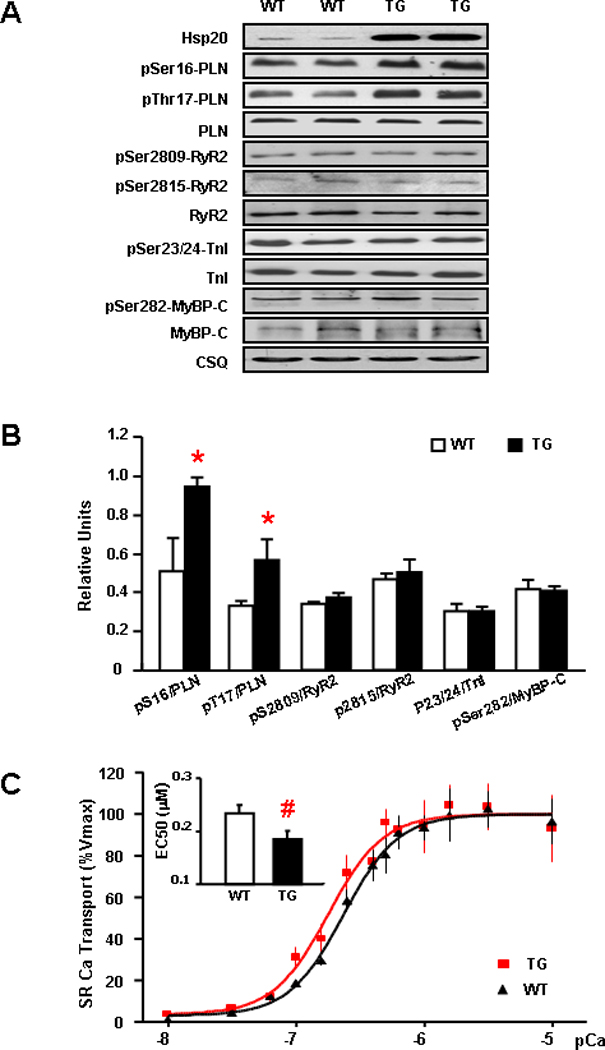
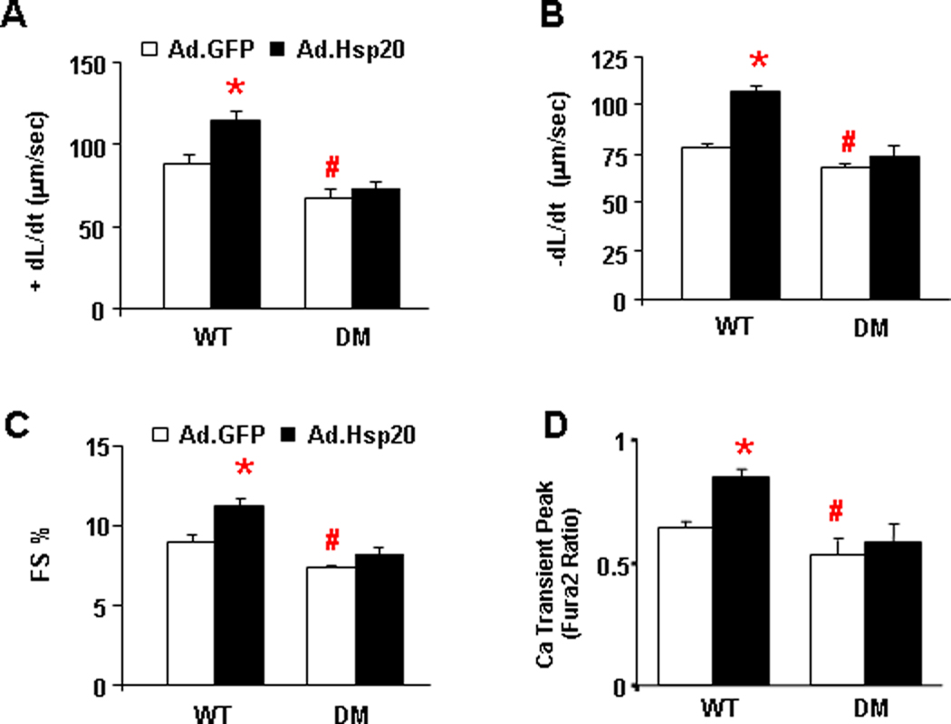
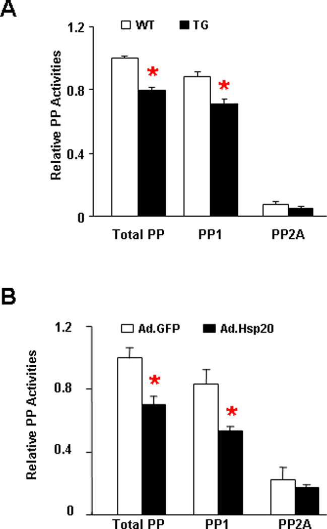
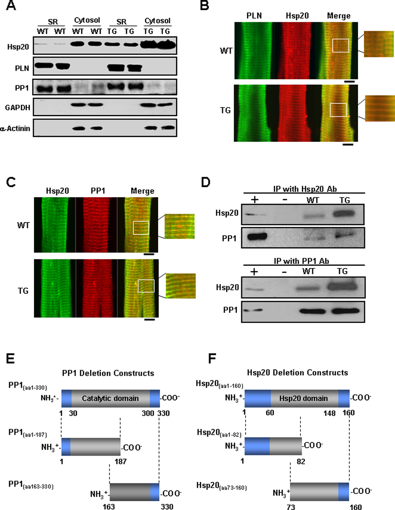
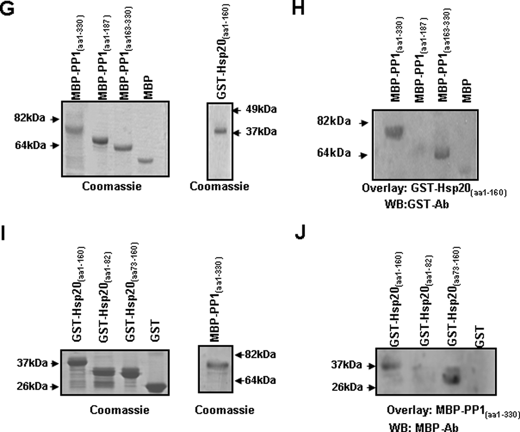
Methods and results: Hsp20 overexpression in intact animals resulted in significant enhancement of cardiac function, coupled with augmented Ca cycling and sarcoplasmic reticulum Ca load in isolated cardiomyocytes. This was associated with specific increases in phosphorylation of phospholamban (PLN) at both Ser16 and Thr17, relieving its inhibition of the apparent Ca affinity of SERCA2a. Accordingly, the inotropic effects of Hsp20 were abrogated in cardiomyocytes expressing nonphosphorylatable PLN (S16A/T17A). Interestingly, the activity of type 1 protein phosphatase (PP1), a known regulator of PLN signaling, was significantly reduced by Hsp20 overexpression, suggesting that the Hsp20 stimulatory effects are partially mediated through the PP1-PLN axis. This hypothesis was supported by cell fractionation, coimmunoprecipitation, and coimmunolocalization studies, which revealed an association between Hsp20, PP1, and PLN. Furthermore, recombinant protein studies confirmed a physical interaction between AA 73 to 160 in Hsp20 and AA 163 to 330 in PP1.

Conclusions: Hsp20 is a novel regulator of sarcoplasmic reticulum Ca cycling by targeting the PP1-PLN axis. These findings, coupled with the well-recognized cardioprotective role of Hsp20, suggest a dual benefit of targeting Hsp20 in heart disease.


Figures

Figure 1
Figure 1. Mechanics and Ca transients of isolated wild type and Hsp20 TG cardiomyocytes
Isolated cardiomyocytes from 12–16 weeks old mice were suspended in 1.8mM Ca-Tyrode solution and field-stimulated at 0.5Hz. (A) Representative cell shortening traces of WT and TG cells. (B) Maximum rates of contraction (+dL/dt). (C) Maximum rates of relaxation (−dL/dt). (D) Fractional shortening (FS%) and (E) Representative tracings of Ca transients in WT and TG cardiomyocytes. (F) Ca transient peak. (G) Time to 50% of decay of Ca transient in WT and Hsp20 TG myocytes. (H) Representative tracings of caffeine induced Ca transient. (I) Caffeine-induced Ca transient amplitude and (J) Time to 50% decay of caffeine-induced Ca transient peak. n = 36–42 cells from 5 hearts for each group. Values = mean ± SEM. *: P<0.01 vs. WT.
Figure 2
Figure 2. SR Ca regulatory proteins in Hsp20 TG hearts
(A) Representative blots of SR Ca-cycling proteins in WT and Hsp20 TG hearts. (B) Quantitative results revealed that SERCA2a, phospholamban pentamer (PLNp), phospholamban monomer (PLNm), total PLN, ryanodine receptor (RYR2), calsequestrin (CSQ), sodium/calcium exchanger (NCX) and L-type Ca channel (LTCC) levels were not altered. (n=6 for each protein). (C) Typical L-type Ca currents in WT and TG cardiomyocytes elicited by a series of depolarizing steps from a holding potential of −50 mV. (D) Average peak current density-voltage relationships of L-type Ca currents recorded in WT (n=15) and TG (n=12) cardiomyocytes. (E) Ramp voltage protocol (top panel) is used to elicit membrane currents (middle panel) in absence and presence of 5mM NiCl2, and the subtraction of the 2 traces gives the Ni2+-sensitive NCX current (bottom panel). (F) Average current density-voltage relationships of NCX currents recorded in WT (n=10) and TG (n=11) ventricular myocytes.
Figure 3
Figure 3. Overexpression of Hsp20 facilitates SR Ca-cycling by phosphorylation of PLN
(A) Representative blots of phosphorylation and total levels of PLN, RyR2, TnI and MyBP-C in WT and Hsp20 TG hearts. (B) Quantitative results of phosphorylated Ca regulatory proteins. Immunoblots revealed that pSer16-PLN/PLN and pThr17-PLN/PLN were significantly increased in TGs compared with wild types, but the phosphorylation levels of Ser2809-RYR2, Ser2815-RyR2, Ser23/24-TnI and Ser282-MyBP-C were not different between WTs and TGs. n=6 for each protein. Values = mean ± SEM. *: P<0.01. (C) Initial rates of oxalate-supported SR Ca-uptake in hearts from WT (▲) and Hsp20 TG (■) mice. There was no difference in the maximal SR Ca- uptake rates, but the EC50 value significantly decreased (inset) in TG hearts, compared to WTs. n= 5 for each group; experiments were performed in duplicates. Values = mean ± SEM, #: P < 0.05.
Figure 4
Figure 4. The inotropic effects of Hsp20 are abrogated in the presence of non-phosphorylatable PLN
(A) Maximum rates of contraction (+dL/dt), (B) maximum rates of relaxation (−dL/dt), (C) fractional shortening (FS%) and (D) twitch Ca amplitude in cultured WT and PLN double-mutant (DM: S16A/T17A) cardiomyocytes upon Ad.GFP and Ad.Hsp20 infection. n=20 cells for WT and n=22 cells for DM. Values = mean ± SEM. *:P<0.05. WT-Ad.Hsp20 vs. WT- Ad.GFP; #: P<0.05. DM-Ad.GFP vs. WT-Ad.GFP.
Figure 5
Figure 5. Hsp20 decreases PP1 activity
(A) Total, PP1 and PP2A activity were measured in the homogenate from the LV myocardium of wild type and Hsp20 transgenic hearts, which were normalized to total phosphatase activity in WT (n=9). Values=mean ± SEM, *:P <0.01 vs. wild type. (B) Total, PP1 and PP2A activity in adenoviral GFP and Hsp20 infected adult rat cardiomyocytes, normalized to total phosphatase activity in Ad.GFP group (n=6). Values= mean ± SEM. *: P<0.01 vs. Ad.GFP, #: P<0.05 vs. Ad.Hsp20.
Figure 6
Figure 6. Interaction of Hsp20 with PP1
(A) Representative blots of Hsp20, PLN, PP1, GAPDH and α-actinin in microsomes enriched in SR membranes or in cytosolic fractions from WT and Hsp20 TG hearts (n=6). The yield of cardiac SR and cytosolic proteins averaged 1.5mg/heart and 12mg/ heart, respectively. The amount of total protein on each gel lane was the same (20 µg for each fraction), which diluted the cytosolic fraction by 10× compared to the SR fraction. (B) WT and TG cardiomyocytes were immunostained for Hsp20 (red) in combination with PLN (green). (C) WT and TG cardiomyocytes were immunostained for Hsp20 (green) in combination with PP1 (red). Scale bar, 10 µm. (D) Co-immunoprecipitation was performed using anti-Hsp20 or anti-PP1 antibody and cardiac homogenates (200µg total protein) of wild type and Hsp20 TG mice. The precipitates were analyzed by immunoblotting with anti-Hsp20 or anti-PP1 antibodies, as indicated. Preimmunoprecipitated WT heart homogenate was used as positive control (+), and immunoprecipitate with anti-IgG PLUS agarose was used as negative control (−). IP: immunoprecipitation. (E) Diagrammatic representation of the full length and the two deletion constructs of PP1 and (F) the full length and the two deletion constructs of Hsp20. Predicted protein domains are shown in grey. (G) SDS-gel stained with Coomassie blue showing the purified MBP-PP1 full-length or deletion proteins. (H) Blot overlay assays with anti-GST antibody (WB : GST-Ab) were performed to determine the protein region of PP1 required for its association with GST-Hsp20(aa1–160). This narrowed down the PP1 binding region to a C-terminal fragment including amino acids 163–330. (I) SDS-gel stained with Coomassie blue showing the purified GST-Hsp20 full-length or deletion proteins. (J) Blot overlay assays with anti-MBP antibody (WB: MBP-Ab) determined that the protein region of Hsp20 responsible for its binding with MBP-PP1(aa1–330) includes amino acids 73–160. WB: Western blot.
Figure 7
Figure 7. Proposed Mechanism of Hsp20 Regulating Cardiac Contractility
The intricate balance between protein kinase and phosphatase activities regulates the phosphorylation of phospholamban in cardiomyocytes. Dephosphorylated PLN poses an inhibitory effect on the activity of SERCA. Dephosphorylation of PLN is mainly mediated by PP1. PKA- and CaMKII-dependent phosphorylation of PLN at Ser16 and Thr17, respectively, relieves PLN inhibition of SERCA and allows for increased Ca-pumping into the SR. Through inhibition of PP1 activity, overexpression of Hsp20 shifts the balance of kinase and PP1 activities, and favors enhanced phosphorylation of PLN and increased cardiac performance.

References

    1. Dreiza CM, Komalavilas P, Furnish EJ, Flynn CR, Sheller MR, Smoke CC, Lopes LB, Brophy CM. The small heat shock protein, HSPB6, in muscle function and disease. Cell Stress Chaperones. 2010;15:1–11. - PMC - PubMed
    1. Qian J, Ren X, Wang X, Zhang P, Jones WK, Molkentin JD, Fan GC, Kranias EG. Blockade of Hsp20 phosphorylation exacerbates cardiac ischemia/reperfusion injury by suppressed autophagy and increased cell death. Circ Res. 2009;105:1223–1231. - PMC - PubMed
    1. Boluyt M, Brevick J, Rogers D, Randall M, Scalia A, Li Z. Changes in the rat heart proteome induced by exercise training: Increased abundance of heat shock protein hsp20. Proteomics. 2006;6:3154–3169. - PubMed
    1. Dohke T, Wada A, Isono T, Fujii M, Yamamoto T, Tsutamoto T, Horie M. Proteomic analysis reveals significant alternations of cardiac small heat shock protein expression in congestive heart failure. J Card Fail. 2006;12:77–84. - PubMed
    1. Fan G, Zhou X, Wang X, Song G, Qian J, Nicolaou P, Chen G, Ren X, Kranias E. Heat shock protein 20 interacting with phosphorylated Akt reduces doxorubicin-triggered oxidative stress and cardiotoxicity. Circ Res. 2008;103:1270–1279. - PMC - PubMed

Publication types

MeSH terms