. 2011 May 27;108(11):1316-27.
doi: 10.1161/CIRCRESAHA.111.243758. Epub 2011 Apr 14.

Mast cell tryptase deficiency attenuates mouse abdominal aortic aneurysm formation

Affiliations

Mast cell tryptase deficiency attenuates mouse abdominal aortic aneurysm formation

Jie Zhang et al. Circ Res. .

Abstract

Rationale: Mast cells (MCs) contribute to the formation of abdominal aortic aneurysms (AAAs) by producing biologically active mediators. Tryptase is the most abundant MC granule protein and participates in MC activation, protease maturation, leukocyte recruitment, and angiogenesis-all processes critical to AAA pathogenesis.

Objective: To test the hypothesis that tryptase participates directly in AAA formation.

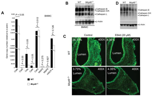
Methods and results: Immunohistochemistry demonstrated enhanced tryptase staining in media and adventitia of human and mouse AAA lesions. Serum tryptase levels correlated significantly with the annual expansion rate of AAA before (r = 0.30, P = 0.003) and after (r = 0.29, P = 0.005) adjustment for common AAA risk factors in a patient follow-up study, and associated with risks for later surgical repair or overall mortality before (P = 0.009, P = 0.065) and after (P = 0.004, P = 0.001) the adjustment. Using MC protease-6-deficient mice (Mcpt6(-/-)) and aortic elastase perfusion-induced experimental AAAs, we proved a direct role of this tryptase in AAA pathogenesis. Whereas all wild-type (WT) mice developed AAA at 14 or 56 days postperfusion, Mcpt6(-/-) mice were fully protected. AAA lesions from Mcpt6(-/-) mice had fewer inflammatory and apoptotic cells, and lower chemokine levels, than did those from WT mice. MC from WT mice restored reduced AAA lesions and lesion inflammatory cell content in MC-deficient Kit(W-sh/W-sh) mice, but those prepared from Mcpt6(-/-) mice did not. Mechanistic studies demonstrated that tryptase deficiency affected endothelial cell (EC) chemokine and cytokine expression, monocyte transmigration, smooth-muscle cell apoptosis, and MC and AAA lesion cysteinyl cathepsin expression and activities.

Conclusions: This study establishes the direct participation of MC tryptase in the pathogenesis of experimental AAAs, and suggests that levels of this protease can serve as a novel biomarker for abdominal aortic expansion.


Figures

Figure 1
Figure 1
Tryptase in human and mouse AAA. A. Correlation of serum tryptase level with annual AAA expansion rate, before (r=0.30, P=0.003) or after (r=0.29, P=0.005) adjustment for AAA confounders, Pearson's correlation test. B. Tryptase and elastin immunostaining in normal (left two panels) and aneurysmal (right two panels) human aortas. Bar: 400 μm; in inset (bottom panels), bar: 100 μm. C. Human AAA and normal aortic tissue extract immunoblot for human tryptase. Tryptase monomer (30-kDa) is indicated. Beta-actin serves as a protein loading control. D. Mouse mMCP-6 polyclonal antibody immunostaining in frozen sections from normal mouse aorta, AAA lesions from Mcpt6−/− (as negative control) and WT mice. Lumen, media, and adventitia are indicated. Bar: 100 μm.
Figure 2
Figure 2
Characterizations of AAA lesions from WT and Mcpt6−/− mice. A. Aortic diameter increase (%). B. Mac-3+ macrophage area (%). C. CD3+ T cell numbers. D. Alpha-actin–positive SMC grade. E. MHC class II-positive area (%). F. MCP-1–positive area (%). Representative images for panels B, C, E, F, H, and I are shown on the right. The number of mice in each experimental group is shown within each bar. Data are mean ± SEM. P<0.05 was considered statistically significant, Mann-Whitney U test.
Figure 3
Figure 3
MC reconstitution in KitW-sh/W-sh mice. WT, MC-deficient KitW-sh/W-sh mice, and those receiving BMMC from WT and Mcpt6−/− mice were induced to produce AAA. Aortic diameter increase (%) (A), Mac-3+ macrophage areas (B), CD3+ T cell numbers (C), and CD31+ microvessel numbers (D) were analyzed at 14 days and 56 days post-perfusion. Numbers of mice for each experimental group are shown in each bar. Data are mean ± SEM. P<0.008 was considered statistically significant, Mann-Whitney U test.
Figure 4
Figure 4
EC cytokine and chemokine expression, monocyte migration, and SMC apoptosis. A. RT-PCR analysis of cytokine and chemokine expression in mouse aortic EC after incubation with degranulated BMMC from WT and Mcpt6−/− mice. Data are mean ± SE of three experiments. B. Peripheral monocyte transmigration assay under different concentrations of SDF-1a. Data are mean ± SE of six experiments. C. RT-PCR determines expression of cathepsins B, S, L, and K in peripheral blood monocytes from WT and Mcpt6−/− mice. Data are mean ± SE of three experiments. P<0.05 was considered statistically significant, Mann-Whitney U test. D. Mouse aortic SMC apoptosis after induction with PDTC with or without BMMC from WT or Mcpt6−/− mice. Representative panels are presented to the left. Green fluorescent cells indicate TUNEL-positive apoptotic cells. P<0.02 was considered statistically significant, Mann-Whitney U test.
Figure 5
Figure 5
Cysteine protease cathepsin expression and activity in MCs and AAA lesions. A. RT-PCR to assess the expression of common MC proteases in BMMC from WT and Mcpt6−/− mice. Data are mean ± SE of three experiments. P<0.05 was considered statistically significant, Mann-Whitney U test. B. Cysteine protease cathepsin active site JPM-labeling with cell lysate from WT and Mcpt6−/− BMMC. C. WT and Mcpt6−/− mouse frozen AAA cross section in situ elastase activity zymograph in the presence or absence of cathepsin inhibitor E64d. Lumen and percentage of fluorescence intensity are indicated. Images were obtained with the same magnification and shutter speed, and all data are from the 56-day time point experiments. D. Cysteine protease cathepsin active site JPM-labeling with aortic tissue lysates from WT and Mcpt6−/− mice from the 56-day time point experiments. In panels B and D, active cathepsins B, S, K, and L are indicated with arrowheads. ß-actin blot was used to ensure equal protein loading.

References

    1. Miller JS, Moxley G, Schwartz LB. Cloning and characterization of a second complementary DNA for human tryptase. J Clin Invest. 1990;86:864–870. - PMC - PubMed
    1. Schwartz LB, Sakai K, Bradford TR, Ren S, Zweiman B, Worobec AS, Metcalfe DD. The alpha form of human tryptase is the predominant type present in blood at baseline in normal subjects and is elevated in those with systemic mastocytosis. J Clin Invest. 1995;96:2702–2710. - PMC - PubMed
    1. Reynolds DS, Gurley DS, Austen KF, Serafin WE. Cloning of the cDNA and gene of mouse mast cell protease-6. Transcription by progenitor mast cells and mast cells of the connective tissue subclass. J Biol Chem. 1991;266:3847–3853. - PubMed
    1. Cairns JA, Walls AF. Mast cell tryptase is a mitogen for epithelial cells. Stimulation of IL-8 production and intercellular adhesion molecule-1 expression. J Immunol. 1996;156:275–283. - PubMed
    1. He S, Peng Q, Walls AF. Potent induction of a neutrophil and eosinophil-rich infiltrate in vivo by human mast cell tryptase: selective enhancement of eosinophil recruitment by histamine. J Immunol. 1997;159:6216–6225. - PubMed

Publication types

MeSH terms