Comparative Study
. 2011 Jun;4(3):344-51.
doi: 10.1161/CIRCEP.110.959312. Epub 2011 Apr 14.

Characterization of gap junction remodeling in epicardial border zone of healing canine infarcts and electrophysiological effects of partial reversal by rotigaptide

Affiliations
Comparative Study

Characterization of gap junction remodeling in epicardial border zone of healing canine infarcts and electrophysiological effects of partial reversal by rotigaptide

Ester Macia et al. Circ Arrhythm Electrophysiol. 2011 Jun.

Abstract

Background: The border zone of healing myocardial infarcts is an arrhythmogenic substrate, partly the result of structural and functional remodeling of the ventricular gap junction protein, Connexin43 (Cx43). Cx43 in arrhythmogenic substrates is a potential target for antiarrhythmic therapy.

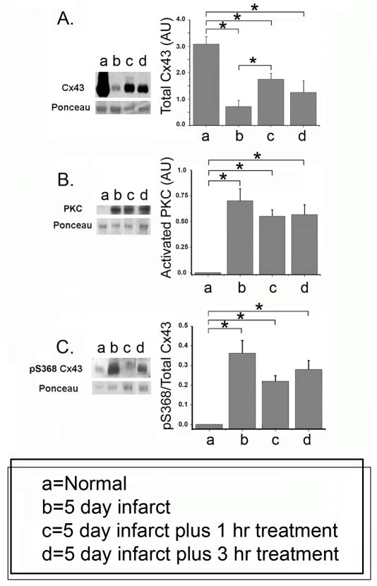
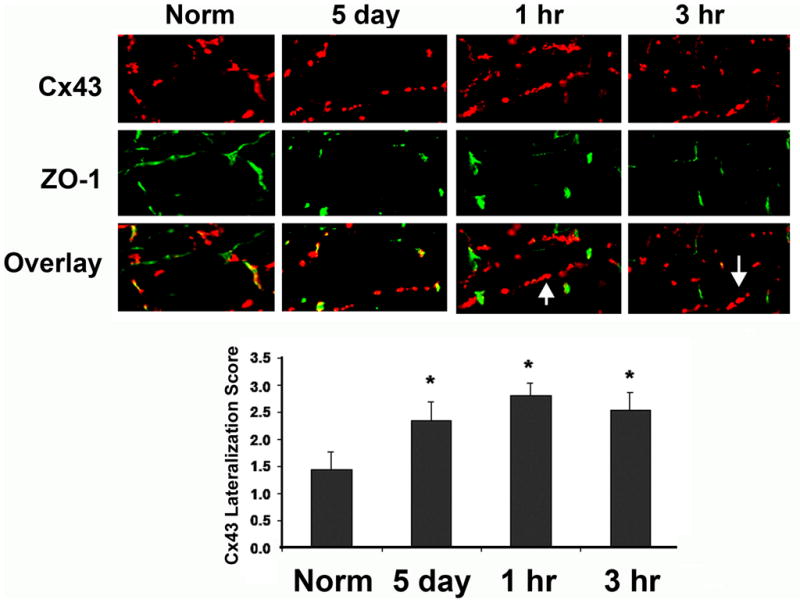
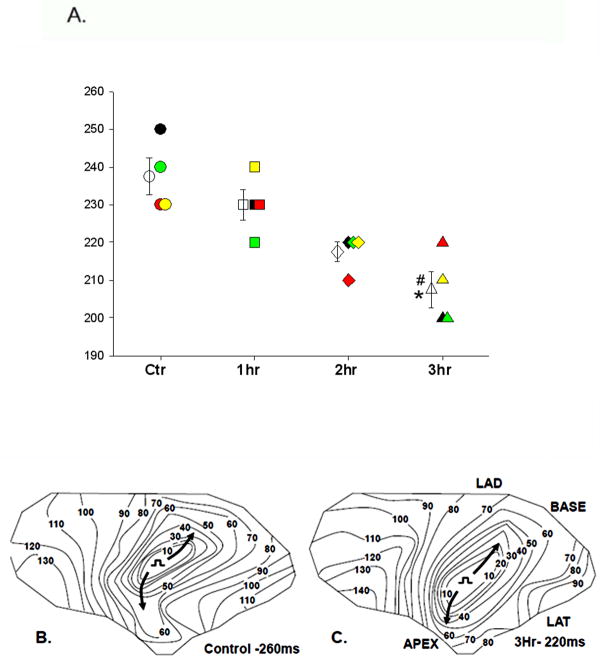
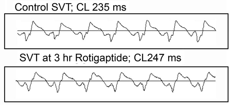
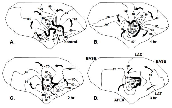
Methods and results: We characterized Cx43 remodeling in the epicardial border zone (EBZ) of healing canine infarcts 5 days after coronary occlusion and examined whether the gap junction-specific agent rotigaptide could reverse it. Cx43 remodeling in the EBZ was characterized by a decrease in Cx43 protein, lateralization, and increased Cx43 phosphorylation at serine (S) 368. Rotigaptide partially reversed the loss of Cx43 but did not affect the increase in S368 phosphorylation, nor did it reverse Cx43 lateralization. Rotigaptide did not prevent conduction slowing in the EBZ, nor did it decrease the induction of sustained ventricular tachycardia by programmed stimulation, although it did decrease the EBZ effective refractory period.

Conclusions: We conclude that partial reversal of Cx43 remodeling in healing infarct border zone may not be sufficient to restore normal conduction or prevent arrhythmias.


Conflict of interest statement

Conflict of Interest Disclosures: This work was supported in part by a grant from Wyeth Pharmaceuticals who supplied the Rotigaptide for these studies (ALW).

Figures

Figure 1
Figure 1
Cx43 protein (A), PKC (B) and Cx43 pS368 (C) in EBZ and effects of Rotigaptide. Representative Western blots are shown at the left. Data (mean values +/− S.E. n=) are shown for normal hearts (no infarct), 5 day infarcts with saline only infusion (b), 5 day infarct with either 1 (c) or 3 (d) hour Rotigaptide infusion. Statistically significant changes indicated by horizontal lines with asterisks above each panel (N=4, p<0.05).
Figure 2
Figure 2
Immunofluorescent labeled myocytes for Cx43 (red) and ZO-1 (green). In normal hearts Cx43 is localized almost exclusively with ZO-1 at the intercalated disk (Norm) (overlay). After 5 day infarction lateralization of Cx43 is seen. Treatment with 1 hr or 3 hr Rotigaptide did not alter lateralization (arrows show areas of lateralized Cx43). Quantification of Cx43 on lateral membranes (not associated with ZO-1) showed no significant effect of Rotigaptide on lateralized Cx43, (N=3, Bar graphs below, mean values +/− S.E).
Figure 3
Figure 3
Activation maps from stimulation of EBZ at the center of the large electrode array in A and B. 10 msec interval isochrones are represented by colors progressing from earliest activation at the site of stimulation (red) to latest activation (dark blue). Panel A shows a control map at a basic cycle length (S1-S1) of 300ms (CVL = 48 cm/s, CVT = 33 cm/s, AR = 1.39) and panel B after 3 hour infusion of Rotigaptide (CVL = 40 cm/s, CVT = 25 cm/s, AR = 1.68). Panels C and D show activation maps with high density array during stimulation from the center at a CL of 300ms. Color code is the same as in Panels A and B, small numbers indicate activation times. The long axes of the elliptical isochrones indicate longitudinal activation (CVL) while the short axes indicate transverse activation (CVT). Panel C shows activation map during control (CVL = 35 cm/s, CVT = 15 cm/s, AR = 2.19), Panel D shows activation map after 3 hours of Rotigaptide (CVL = 33 cm/s, CVT = 16 cm/s, AR = 2.18).
Figure 4
Figure 4
Panel A. Panel A. Effective refractory periods (EBZ-RP) in control before Rotigaptide infusion (Ctr circles;N=4), and 1 hour (1hr squares; N=4), 2 (2hr diamonds; N=4) and 3 (3hr triangles; N=4) after infusion. Data from each individual experiment are represented by a different color (black, red, yellow and green). Mean±SE at each time is represented by the unfilled symbols with error bars. *P<0.05 3hr vs Ctr and #P<0.05 3hr vs 1hr determined by repeated measures ANOVA/Bonferroni’s. Panel B shows activation of the earliest propagated premature impulse in control (S1–S2= 260 ms) and Panel C shows the activation of the earliest propagated premature impulse after 3 hours Rotigaptide infusion (S1–S2 = 220 ms), when ERP was decreased. Longitudinal activation (arrows) is similar before and after Rotigaptide.
Figure 5
Figure 5
ECG recorded during control SMVT (top panel) and after 3 hour Rotigaptide infusion.
Figure 6
Figure 6
Effects of Rotigaptide on a reentrant circuit causing SMVT. Each panel shows an activation map of the EBZ, isochrones at 10 msec intervals, arrows indicate direction of activation. Dark lines indicate regions of block. Panel A shows activation pattern in control. The pattern is a figure of eight reentrant circuit with a central common pathway between the two lines of block. In this central region of the EBZ there is no indication of transmural break through although at the periphery where the EBZ abuts non infarcted myocardium (within 100 msec isochrones) there is likely transmural activation. Panel B shows the activation pattern after 1 hour Rotigaptide infusion. The lines of block have shifted and the reentrant cycle length is prolonged because the direction of activation in the central common pathway has shifted from longitudinal, to transverse. Panel C shows the pattern of activation during the SVT induced after 2 hours of Rotigaptide infusion. The pattern is similar to control with minor shifts in block lines. Panel D shows activation of SVT after 3 hours of Rotigaptide infusion. A reentrant circuit is no longer mapped in the EBZ. The border zone is activated nearly simultaneously by multiple wave fronts moving from the margins toward the center. There is transmural breakthrough around the entire margin of the electrode array which overlies non infarcted myocardium.

Similar articles

Cited by

References

    1. Dillon S, Allessie M, Ursell PC, Wit AL. Influence of anisotropic tissue structure on reentrant circuits in the subepicardial border zone of subacute canine infarcts. Circulation Research. 1988;63:182–206. - PubMed
    1. Peters NS, Coromilas J, Severs NJ, Wit AL. Disturbed connexin43 gap junction distribution correlates with the location of reentrant circuits in the epicardial border zone of healing canine infarcts that cause ventricular tachycardia. Circulation. 1997;95:988–996. - PubMed
    1. Severs NJ, Bruce AF, Dupont E, Rothery S. Remodeling of gap junctions and connexin expression in diseased myocardium. Cardiovascular Research. 2008;80:9–19. - PMC - PubMed
    1. Lampe PD, Cooper CD, King TJ, Burt JM. Analysis of connexin43 phosphorylated at S325, S328 and S330 in normoxic and ischemic heart. J Cell Sci. 2006;119:3435–3442. - PMC - PubMed
    1. Solan JL, Lampe PD. Key Connexin 43 Phosphorylation Events Regulate the Gap Junction Life Cycle. J Membrane Biol. 2007;217:35–41. - PMC - PubMed

Publication types

MeSH terms