

Vogt-Koyanagi-Harada disease occurring during pegylated interferon-α2b and ribavirin combination therapy for chronic hepatitis C
- PMID: 21494079
- PMCID: PMC3304626
- DOI: 10.3350/kjhep.2011.17.1.61
Vogt-Koyanagi-Harada disease occurring during pegylated interferon-α2b and ribavirin combination therapy for chronic hepatitis C
Abstract
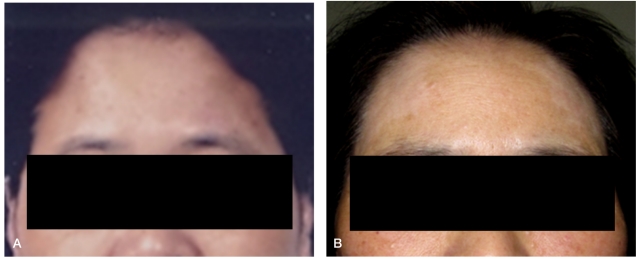
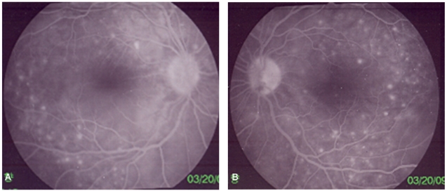
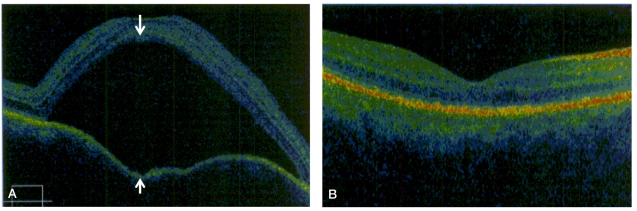
Vogt-Koyanagi-Harada (VKH) disease is a multisystem syndrome characterized by ocular (uveitis and retinal detachment), neurological (headache, tinnitus, and meningitis), and integumentary (vitiligo, alopecia, and poliosis) involvement. Although the pathogenesis of VKH disease is not well understood, an autoimmune T-cell response to a melanocyte-associated antigen is considered to be a cause of VKH disease. The complex immunological response to interferon and ribavirin may induce or exacerbate the autoimmune condition; however, VKH disease is a very rare complication associated with interferon therapy in chronic hepatitis C. We report a case of VKH disease occurring during pegylated interferon-α2b and ribavirin combination therapy for chronic hepatitis C.
Figures
References
-
- Fattovich G, Giustina G, Favarato S, Ruol A. A survey of adverse events in 11,241 patients with chronic viral hepatitis treated with alfa interferon. J Hepatol. 1996;24:38–47. - PubMed
-
- Kasahara A, Hiraide A, Tomita N, Iwahashi H, Imagawa A, Ohguro N, et al. Vogt-Koyanagi-Harada disease occurring during interferon alpha therapy for chronic hepatitis C. J Gastroenterol. 2004;39:1106–1109. - PubMed
-
- Vancoillie G, Lambert J, Nayaert JM. Melanocyte biology and its implications for the clinician. Eur J Dermatol. 1999;9:241–251. - PubMed
-
- Kawano T, Shigehira M, Uto H, Nakama T, Kato J, Hayashi K, et al. Retinal complications during interferon therapy for chronic hepatitis C. Am J Gastroenterol. 1996;91:309–313. - PubMed
Publication types
MeSH terms
Substances
LinkOut - more resources
Full Text Sources