

Case Reports
doi: 10.3350/kjhep.2011.17.1.76.
IgG4-related sclerosing cholangitis: liver biopsy findings
Affiliations
- PMID: 21494082
- PMCID: PMC3304628
- DOI: 10.3350/kjhep.2011.17.1.76
Item in Clipboard
Case Reports
IgG4-related sclerosing cholangitis: liver biopsy findings
Korean J Hepatol.
2011 Mar.
No abstract available
Figures
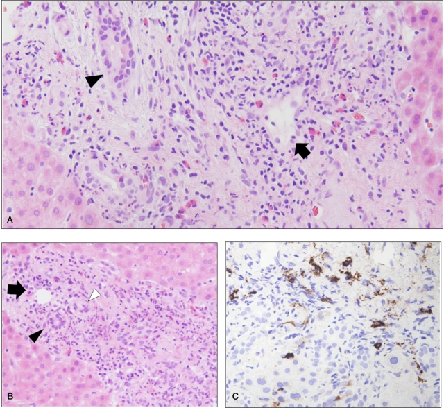
Histologic features of the biopsied liver. (A, B) Lymphoplasmacytic and eosinophilic infiltration in the portal tracts (A: H&E, ×200; B: H&E, ×100). The inflammatory cell infiltrates are centered predominantly around the portal veins (arrows), while the hepatic artery (white arrowhead) and bile duct epithelia (black arrowhead) are relatively intact without degenerative changes. The portal tracts are mildly fibrotic, without an 'onion-skin' appearance. Immunohistochemical stain for IgG4 reveals several scattered IgG4-positive plasma cells in the portal tracts (C: IgG4 immunostaining, ×400).
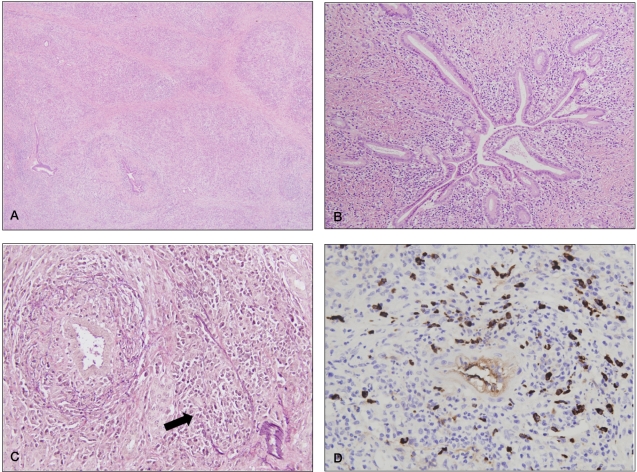
Histologic features of the pancreas (A-C). Diffuse lymphoplasmacytic infiltration of the pancreatic lobules and periductal areas with marked septal fibrosis is seen (A: H&E, ×40). The inflammatory cells infiltrate the periductal areas without destruction of the ductal epithelia (B: H&E, ×100). Obliterative phlebitis is frequently seen (arrow), while ductal epithelia and arteries are spared. (C: Elastic van Gieson, ×200). Abundant IgG4-positive plasma cells are seen in the pancreatic parenchyma (D: IgG4 immunostaining, ×400).
References
-
- Kamisawa T, Funata N, Hayashi Y, Eishi Y, Koike M, Tsuruta K, et al. A new clinicopathological entity of IgG4-related autoimmune disease. J Gastroenterol. 2003;38:982–984. - PubMed
-
- Nakazawa T, Ohara H, Sano H, Ando T, Aoki S, Kobayashi S, et al. Clinical differences between primary sclerosing cholangitis and sclerosing cholangitis with autoimmune pancreatitis. Pancreas. 2005;30:20–25. - PubMed
-
- Nakazawa T, Ando T, Hayashi K, Naitoh I, Ohara H, Joh T. Diagnostic procedures for IgG4-related sclerosing cholangitis. J Hepatobiliary Pancreat Sci. 2010 Sep 02; [Epub ahead of print] - PubMed
-
- Hamano H, Kawa S, Horiuchi A, Unno H, Furuya N, Akamatsu T, et al. High serum IgG4 concentrations in patients with sclerosing pancreatitis. N Engl J Med. 2001;344:732–738. - PubMed
-
- Björnsson E, Chari ST, Smyrk TC, Lindor K. Immunoglobulin G4 associated cholangitis: description of an emerging clinical entity based on review of the literature. Hepatology. 2007;45:1547–1554. - PubMed
Publication types
MeSH terms
Substances
LinkOut - more resources
Full Text Sources