. 2011 Aug;25(8):1286-1296.
doi: 10.1038/leu.2011.62. Epub 2011 Apr 15.

Rapid mobilization of hematopoietic progenitors by AMD3100 and catecholamines is mediated by CXCR4-dependent SDF-1 release from bone marrow stromal cells

Affiliations

Rapid mobilization of hematopoietic progenitors by AMD3100 and catecholamines is mediated by CXCR4-dependent SDF-1 release from bone marrow stromal cells

Ayelet Dar et al. Leukemia. 2011 Aug.

Erratum in

  • Leukemia. 2011 Aug;25(8):1378

Abstract

Steady-state egress of hematopoietic progenitor cells can be rapidly amplified by mobilizing agents such as AMD3100, the mechanism, however, is poorly understood. We report that AMD3100 increased the homeostatic release of the chemokine stromal cell derived factor-1 (SDF-1) to the circulation in mice and non-human primates. Neutralizing antibodies against CXCR4 or SDF-1 inhibited both steady state and AMD3100-induced SDF-1 release and reduced egress of murine progenitor cells over mature leukocytes. Intra-bone injection of biotinylated SDF-1 also enhanced release of this chemokine and murine progenitor cell mobilization. AMD3100 directly induced SDF-1 release from CXCR4(+) human bone marrow osteoblasts and endothelial cells and activated uPA in a CXCR4/JNK-dependent manner. Additionally, ROS inhibition reduced AMD3100-induced SDF-1 release, activation of circulating uPA and mobilization of progenitor cells. Norepinephrine treatment, mimicking acute stress, rapidly increased SDF-1 release and progenitor cell mobilization, whereas β2-adrenergic antagonist inhibited both steady state and AMD3100-induced SDF-1 release and progenitor cell mobilization in mice. In conclusion, this study reveals that SDF-1 release from bone marrow stromal cells to the circulation emerges as a pivotal mechanism essential for steady-state egress and rapid mobilization of hematopoietic progenitor cells, but not mature leukocytes.


Figures
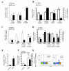
Figure 1
Figure 1
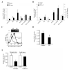
CXCR4-dependent steady state and AMD3100-induced release of stromal derived factor-1 (SDF-1) mediates preferential egress and rapid mobilization of progenitor cells. (A) Steady state homeostasis fold change in the levels of N-terminally intact SDF-1 in murine plasma and bone marrow (BM) supernatants upon administration of PBS, isotype control Ab or anti-CXCR4 Ab. Values of plasma SDF-1 levels: 0.7±0.2 ng/ml, 0.8±-0.3 ng/ml and 0.2±-0.2 ng/ml, respectively. (B) Total number of circulating white blood cells (WBC) and the frequency of colony forming progenitors (CFU-C) in the presence of Ab against SDF-1, CXCR4 or isotype control Ab, in steady state. (C and D) AMD3100-induced release of functional SDF-1 in murine plasma and BM sup. (C) and the frequency of circulating murine WBC and CFU-C (D) one hour after administration. Values of plasma SDF-1 levels: 0.61±0.2 ng/ml, 1.19±-0.34 ng/ml, 1.79±-0.03 ng/ml and 1.12±-0.35 ng/ml, respectively. (E-G) AMD3100-induced release of functional SDF-1 in primate plasma (E), and the frequency of circulating primate WBC and CD34+ progenitor cells (F), one hour after administration. Representative flow cytometry analysis one hour after AMD3100 administration (G). *p<0.05 compared to AMD3100 treated mice (D), or to matched controls (A-C, E and F). #p<0.05 compared to AMD3100 treated mice (C). n=3-5 mice or non-human primates per group.
Figure 2
Figure 2
Kinetics of AMD3100-induced functional SDF-1 release and rapid mobilization. (A and B) AMD3100-induced release of SDF-1 in murine plasma and BM sup. (A) and the frequency of circulating murine WBC and CFU-C (B) 10 minutes after administration. Values of plasma SDF-1 levels: 1.2±0.17 ng/ml and 1.3±0.18 ng/ml, respectively. (C and D) Fold changes in SDF-1 levels in murine plasma and BM sup. (C) and the frequency of circulating WBC and CFU-C (D) were determined one hour and 24 hours after AMD3100 administration or following two injections of AMD3100 every hour (AMD 2×1h). Values of plasma SDF-1 levels: 0.76±0.03 ng/ml, 2.1±0.3 ng/ml, 1.2±0.07 and 0.85±0.08 ng/ml, respectively. *p<0.05 compared to matched controls. n=4-5 mice per group.
Figure 3
Figure 3
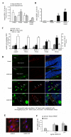
AMD3100 agonistically affects BM stromal cells to release functional SDF-1. (A-C) Mice were injected intra-bone with biotinylated SDF-1 3/6 (bSDF-1) and were either treated with AMD3100 or PBS (s.c). Concentrations of intact intra-femur injected bSDF-1 in BM sup of injected femurs and in non-injected femurs (A) and in the plasma (B) at the indicated time points. Matched control samples from PBS injected mice were negative for the presence of bSDF-1. (C) Total numbers of circulating WBC and the frequency of CFU-C in the plasma and BM sup. n=4-6 mice per group. *p<0.05 compared to control mice; $p<0.05 compared to 30 minutes after bSDF-1 injection; #p<0.05 compared to 60 minutes after bSDF-1 injection (without AMD3100 treatment). (D) Immunofluorescent labeling of the BM of control or AMD3100 treated mice. Immunoreactivity of SDF-1 (red) is observed in PBS treated murine BM: upper panels - osteopontin (green) positive osteoblasts in the bone shaft; lower panels – von Willebrand’s factor (vW, green) positive blood vessels (BV). Blood vessels are marked with arrows or a dashed border. SDF-1 immunoreactivity is not observed in AMD3100 treated murine BM. Nuclear staining by DAPI (Blue). Original magnifications: upper panels – x40; lower panels – x200 (PBS), x630 (AMD3100). (E) Untreated or AMD3100 (100 ng/ml) treated primary human BMEC were immunolabeled for functional SDF-1 (red) and stained for nuclear DNA (purple). (F) Secretion of SDF-1 from cultured BMEC or the osteoblastic human cell line MG-63 in response to AMD3100, measured by ELISA. n=4, *p< 0.05 compared to control.
Figure 4
Figure 4
ROS inhibition reduces AMD3100-induced mobilization and SDF-1 release. (A-B) Mice were treated with AMD3100 or PBS as control as described in Figure 1. In addition, mice were treated with the Reactive Oxygen Species (ROS) inhibitor N-Acetyl Cysteine (NAC) or PBS as control. (A) One hour post injection of AMD3100, circulating WBC (black bars) and CFU-C (white bars) were measured. (B) Functional SDF-1 levels in the plasma. n=4-5 mice per group. Values of plasma SDF-1 levels: 0.71±0.06 ng/ml, 1.72±-0.2 ng/ml, 0.86±-0.12, respectively. *p<0.05 compared to PBS alone and #p<0.05 compared to AMD3100 treatment.
Figure 5
Figure 5
Neurotransmitter stimulation induces functional SDF-1 release and rapid progenitor mobilization. (A-B) SDF-1 levels in the plasma and BM sup. (A) or circulating WBC and progenitor cells (B) in mice treated with norepinephrine (NE) or the β2 adrenergic antagonist ICI, 1 hr after administration. Control mice received injections of PBS. n=6 mice/group. Values of plasma SDF-1 levels: 1.1±0.17 ng/ml, 1.8±0.5 ng/ml, 0.6±0.06, 2±0.3 ng/ml, 2.8±0.5 ng/ml and 1.2±-0.1 ng/ml, respectively. *p<0.05 compared to control mice. #p<0.05 compared to AMD3100 treated mice (C) RT-PCR analysis (top) for mRNA expression and flow cytometry analysis (bottom) for cell surface expression of β2-adrenergic receptor on cultured primary human BMEC. –RT=cDNA was prepared without reverse transcriptase as a control. (D) SDF-1 release from primary human BMEC in response to stimulation with ICI (10 ng/ml). n=3 (E) AMD3100-induced mobilization of progenitors in control and sympathectomised (6OHDA) of either neonate or adult mice. *p<0.005. n=10-15 mice per group.
Figure 6
Figure 6
Rapid mobilization induced by AMD3100 involves JNK and uPA release in a CXCR4-dependent manner. (A) Representative zymography of uPA activity in the plasma of mice treated with either PBS or AMD3100 with or without neutralizing Ab against CXCR4 and matched isotype control, one hour post administration. (B) The frequency of circulating WBC and CFU-C one hour after administration of vehicle or AMD3100 in the presence or absence of JNK inhibitor. *p<0.05 compared to control mice. #p<0.05 compared to AMD3100 treated mice. n=6 mice/group. (C) Activity of uPA in the plasma after AMD3100 administration in the presence or absence of JNK inhibitor. (D) Fold change in the levels of circulating functional SDF-1 one hour after vehicle or AMD3100 administration in the presence or absence of JNK inhibitor. n=6 mice/group. Values of plasma SDF-1 levels: 0.38±0.02 ng/ml, 0.34±0.09 ng/ml, 2±0.04, 1.6±0.3 ng/ml, respectively. *p<0.05. (E) Activity of uPA in the plasma after AMD3100 administration in the presence or absence of ROS inhibitor NAC.

References

    1. Spiegel A, Kalinkovich A, Shivtiel S, Kollet O, Lapidot T. Stem cell regulation via dynamic interactions of the nervous and immune systems with the microenvironment. Cell Stem Cell. 2008;3:484–92. - PubMed
    1. Lapidot T, Kollet O. Hematology. 30th edition 2010. The Brain-Bone-Blood Triad: Traffic Lights for Stem-Cell Homing and Mobilization. - PubMed
    1. Wright DE, Wagers AJ, Gulati AP, Johnson FL, Weissman IL. Physiological migration of hematopoietic stem and progenitor cells. Science. 2001;294:1933–6. - PubMed
    1. Kollet O, Dar A, Lapidot T. The multiple roles of osteoclasts in host defense: bone remodeling and hematopoietic stem cell mobilization. Annu Rev Immunol. 2007;25:51–69. - PubMed
    1. Lapid K, Vagima Y, Kollet O, Lapidot T. Egress and mobilization of hematopoietic progenitor cells. StemBook, Harvard Stem Cell Institute. 2009 1.39.1. - PubMed

Publication types