

Non-dura based intaspinal clear cell meningioma
- PMID: 21494369
- PMCID: PMC3070901
- DOI: 10.3340/jkns.2011.49.1.71
Non-dura based intaspinal clear cell meningioma
Abstract
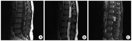
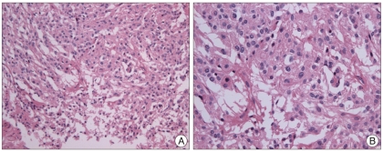
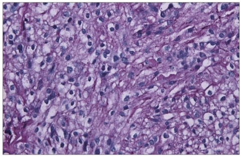
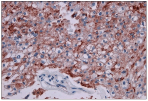
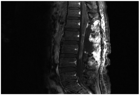
A 34-year-old female patient was presented with leg and hip pain for 6 months as well as voiding difficulty for 1 year. Magnetic resonance imaging revealed a well-demarcated mass lesion at L2-3. The mass was hypo-intense on T1- and T2-weighted images with homogeneous gadolinium enhancement. Surgery was performed with the presumptive diagnosis of intradural extramedullary meningioma. Complete tumor removal was possible due to lack of dural adhesion of the tumor. Histologic diagnosis was clear cell meningioma, a rare and newly included World Health Organization classification of meningioma usually affecting younger patients. During postoperative 2 years, the patient has shown no evidence of recurrence. We report a rare case of cauda equina clear cell meningioma without any dural attachment.
Keywords: Cauda equina; Clear cell meningioma; Spinal meningioma; World Health Organization classification; Younger patients.
Figures
Similar articles
-
Spinal clear cell meningioma without dural attachment: a case report and literature review.Radiol Case Rep. 2022 Mar 25;17(5):1760-1764. doi: 10.1016/j.radcr.2022.02.052. eCollection 2022 May. Radiol Case Rep. 2022. PMID: 35355528 Free PMC article.
-
Lumbar clear cell meningioma mimicking schwannoma 7 years after resection of the same type of intracranial tumor: a case report.J Med Case Rep. 2024 Feb 6;18(1):82. doi: 10.1186/s13256-024-04411-8. J Med Case Rep. 2024. PMID: 38321548 Free PMC article.
-
Giant nondural-based cauda equina meningioma with multiple cysts.J Neurooncol. 2005 Sep;74(2):173-7. doi: 10.1007/s11060-004-2905-6. J Neurooncol. 2005. PMID: 16132526
-
Two cases of nondura-based clear cell meningioma of the cauda equina.APMIS. 2004 Feb;112(2):141-7. doi: 10.1111/j.1600-0463.2004.apm1120209.x. APMIS. 2004. PMID: 15056231 Review.
-
Spinal Meningioma Arising from the Denticulate Ligament.World Neurosurg. 2018 Jul;115:329-333. doi: 10.1016/j.wneu.2018.04.160. Epub 2018 May 3. World Neurosurg. 2018. PMID: 29729464 Review.
Cited by
-
Clear cell meningioma of the lower lumbar spine without dural attachment: A case report.Medicine (Baltimore). 2025 Jul 11;104(28):e43193. doi: 10.1097/MD.0000000000043193. Medicine (Baltimore). 2025. PMID: 40663004 Free PMC article.
-
Atypical meningioma originating from the spinal accessory nerve.Surg Neurol Int. 2022 Dec 31;13:598. doi: 10.25259/SNI_1085_2022. eCollection 2022. Surg Neurol Int. 2022. PMID: 36761262 Free PMC article.
-
A Giant Nondural-Based Lumbosacral Clear Cell Meningioma Mimicking Schwannoma: A Case Report and Review of the Literature.Asian J Neurosurg. 2021 Feb 23;16(1):44-50. doi: 10.4103/ajns.AJNS_385_20. eCollection 2021 Jan-Mar. Asian J Neurosurg. 2021. PMID: 34211865 Free PMC article. Review.
-
Spinal clear cell meningioma without dural attachment: a case report and literature review.Radiol Case Rep. 2022 Mar 25;17(5):1760-1764. doi: 10.1016/j.radcr.2022.02.052. eCollection 2022 May. Radiol Case Rep. 2022. PMID: 35355528 Free PMC article.
-
Loss of SMARCE1 expression is a specific diagnostic marker of clear cell meningioma: a comprehensive immunophenotypical and molecular analysis.Brain Pathol. 2018 Jul;28(4):466-474. doi: 10.1111/bpa.12524. Epub 2017 Jun 12. Brain Pathol. 2018. PMID: 28474749 Free PMC article.
References
-
- Carra S, Drigo P, Gardiman M, Perilongo G, Rigobello L. Clear-cell meningioma in a 22-month-old male : a case report and literature review. Pediatr Neurosurg. 2001;34:264–267. - PubMed
-
- Chen MH, Chen SJ, Lin SM, Chen MH. A lumbar clear cell meningioma with foraminal extension in a renal transplant recipient. J Clin Neurosci. 2004;11:665–667. - PubMed
-
- Dhall SS, Tumialán LM, Brat DJ, Barrow DL. Spinal intradural clear cell meningioma following resection of a suprasellar clear cell meningioma. Case report and recommendations for management. J Neurosurg. 2005;103:559–563. - PubMed
-
- Dubois A, Sevely A, Boetto S, Delisle MB, Manelfe C. Clear-cell meningioma of the cauda equina. Neuroradiology. 1998;40:743–747. - PubMed
-
- Epstein NE, Drexler S, Schneider J. Clear cell meningioma of the cauda equina in an adult: case report and literature review. J Spinal Disord Tech. 2005;18:539–543. - PubMed
Publication types
LinkOut - more resources
Full Text Sources
