

Two sporadic infected cardiac myxomas in 1 patient
- PMID: 21494535
- PMCID: PMC3066802
Two sporadic infected cardiac myxomas in 1 patient
Abstract
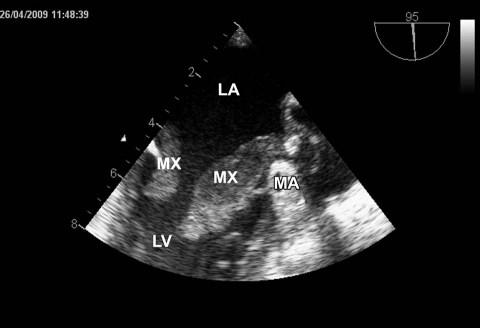
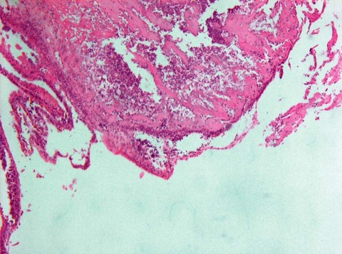
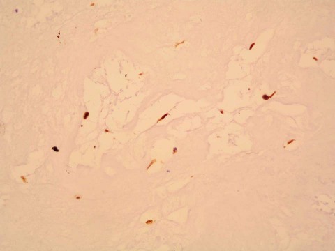
Infected cardiac myxoma is a rare cause of endocarditis. The finding of coexisting infected cardiac myxomas is highly unusual. Herein, we present the case of a 58-year-old woman with a low-grade fever. Laboratory findings strongly indicated inflammation, and blood cultures detected Staphylococcus species. Echocardiograms revealed mobile masses in the area of the mitral valve. Transesophageal echocardiograms showed 2 formations that arose from opposite sides of the mitral annulus and protruded into the left ventricle during systole. During emergency surgery, 2 abnormal growths with numerous vegetations were completely excised. The diagnosis of myxoma was confirmed upon histologic evaluation. Microbiological and polymerase chain reaction analysis of the myxomas detected the bacterial strain Enterococcus faecalis. Five months postoperatively, the patient showed no signs of recurrent infection and had a normal echocardiographic appearance.This report is the first of an infected cardiac myxoma in the Czech population and one of approximately 60 reports in the medical literature from 1956 to the present. In addition to the case of our patient, we discuss the discrepancy between the bacteriologic findings.
Keywords: Bacteroidaceaeinfections/complications/diagnosis/drug therapy; echocardiography; heart atria/pathology; heart neoplasms/diagnosis/surgery; heart valve diseases/surgery; myxoma/diagnosis/pathology/surgery; neoplasms, second primary; treatment outcome.
Figures
References
-
- Schaff HV, Mullany CJ. Surgery for cardiac myxomas. Semin Thorac Cardiovasc Surg 2000;12(2):77–88. - PubMed
-
- Tillmanns H. Clinical aspects of cardiac tumors. Thorac Cardiovasc Surg 1990;38 Suppl 2:152–6. - PubMed
-
- Shinfeld A, Katsumata T, Westaby S. Recurrent cardiac myxoma: seeding or multifocal disease? Ann Thorac Surg 1998; 66(1):285–8. - PubMed
-
- Carney JA. Differences between nonfamilial and familial cardiac myxoma. Am J Surg Pathol 1985;9(1):53–5. - PubMed
Publication types
MeSH terms
Substances
LinkOut - more resources
Full Text Sources