. 2011 Apr 11;6(4):e18412.
doi: 10.1371/journal.pone.0018412.

Microarray analysis uncovers a role for Tip60 in nervous system function and general metabolism

Affiliations

Microarray analysis uncovers a role for Tip60 in nervous system function and general metabolism

Meridith Lorbeck et al. PLoS One. .

Abstract

Background: Tip60 is a key histone acetyltransferase (HAT) enzyme that plays a central role in diverse biological processes critical for general cell function; however, the chromatin-mediated cell-type specific developmental pathways that are dependent exclusively upon the HAT activity of Tip60 remain to be explored.

Methods and findings: Here, we investigate the role of Tip60 HAT activity in transcriptional control during multicellular development in vivo by examining genome-wide changes in gene expression in a Drosophila model system specifically depleted for endogenous dTip60 HAT function.

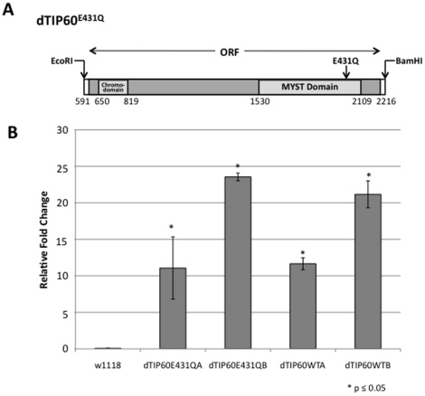
Conclusions: We show that amino acid residue E431 in the catalytic HAT domain of dTip60 is critical for the acetylation of endogenous histone H4 in our fly model in vivo, and demonstrate that dTip60 HAT activity is essential for multicellular development. Moreover, our results uncover a novel role for Tip60 HAT activity in controlling neuronal specific gene expression profiles essential for nervous system function as well as a central regulatory role for Tip60 HAT function in general metabolism.


Conflict of interest statement

Competing Interests: The authors have declared that no competing interests exist.

Figures

Figure 1
Figure 1. Generation and characterization of transgenic dTip60E431Q and dTip60WT flies.
(A) Schematic of the dTIP60 open reading frame. Shown is the location of the conserved regions encoding for the N-terminal chromo domain and the C-terminal MYST functional domain. An arrow denotes the position site of amino acid substitution E431Q. (B) Exogenous expression levels of dTip60E431Q and dTip60WT in independent fly lines. Shown is a histogram depicting qPCR analysis of exogenous levels of dTip60 in staged three day old second instar larvae progeny resulting from a cross between ubiquitous GAL4 drive 337 and either dTip60E431Q (lines A and B), dTip60WT (lines A and B) or control w1118 flies. To quantify the amount of exogenous expressed dTip60E431Q or dTip60WT, primers were designed to amplify a 97bp non-conserved region of dTip60 (present in both endogenous and exogenous dTip60) and a 105bp non-conserved region within the 5′UTR of dTip60, (present only in endogenous dTip60). For each transgenic line, RNA was extracted and qPCR performed using Power Sybr Green. The cycle threshold (CT) was determined for each primer pair and normalized to the CT value of RP49 (ribosomal protein internal loading control) to account for differences between samples. The relative fold change in mRNA expression levels between exogenous and endogenous dTip60 was measured using the comparative Ct method with Rp49 as the internal control, and these results are summarized in the histogram. Asterisks (*) indicate significant fold change between the respective genotype and control flies with values of p≤0.05; n = 3. Error bars represent standard error of the mean.
Figure 2
Figure 2. Expression of dTip60E431Q in flies significantly depletes endogenous levels of histone H4 acetylation in vivo.
(A) Equal amounts of core histones isolated from 50 three day old staged second instar larvae for each genotype crossed to GAL4 line 337 were resolved by 18% polyacrylamide gel electrophoresis, Western-blotted, and immunostained with antibodies that recognize four acetylated lysine residues (K5, K8, K12 and K16) of histone H4. Coomassie staining of core histone proteins were used to ensure equal loading of the samples , . (B) Western blot signals were quantitated using Fluorchem imager (Alpha Innotech) and the results are summarized in the histogram depicting arbitrary units of endogenous histone H4 acetylation for each of the three genotypes analyzed. To ensure signal was in the linear range, Alpha Ease FC software (Alpha Innotech, San Leandro, CA) was used according to the manufacturer's instructions to select exposure times such that there was no saturation detected. Values indicated are the mean of three independent biological replicates. Asterisks (*) indicate significant fold change in acetylation in relation to control w1118 flies where p<0.05. Error bar depicts standard error of the mean; n = 3.
Figure 3
Figure 3. Microarray analysis reveals a central role for Tip60 in the transcriptional control of genes linked to diverse metabolic and general cellular processes.
(A) Total number of significantly misregulated genes in response to dTip60E431Q or dTip60WT. The dCHIP t-test function was used to identify genes whose expression differed significantly (p<0.05) and these genes were then filtered to select for those that showed a twofold or greater change and a 90% confidence bound of fold change. (B) Significantly enriched gene ontology (GO) groups representing dTip60E431Q and dTip60WT misregulated genes. Genes were annotated and biological processes were analyzed using the Database for Annotation, Visualization, and Integrated Discovery (DAVID). Significance of overrepresentation of Gene Ontology (GO) terms was determined (p<0.05). Enrichment score (y-axis) is reported as the minus log transformation on the geometric mean of p-values from the enriched annotation terms associating with one or more of the gene group members. The genes up-regulated in response to dTip60E431Q clustered into 5 significantly enriched groups, and down-regulated genes clustered into 12 significantly enrichment groups, with 8 of these groups enriched for metabolic processes. Genes misregulated in response to dTip60WT grouped to one significantly enriched cluster.
Figure 4
Figure 4. qRT-PCR validation of selected neuronal target genes identified by microarray analysis.
Shown is a histogram depicting qPCR analysis of the expression of selected neuronal target genes identified by microarray using aliquots of cDNA pools prepared for microarray analysis. The relative fold change in mRNA expression levels were measured using the comparative Ct method with RP49 as the internal control gene. Asterisk (*) indicates significant fold change where * is p≤0.05, ** is p≤0.0005 and *** p≤0.000008. Error bars represent standard deviation and are reported as ± standard error of the mean, (n = 3).
Figure 5
Figure 5. Tip60 localization in the nervous system of Drosophila embryos.
Confocal microscopic dorsal view of a w1118 wild-type embryo (stage 15) double labeled with Tip60 antibody (blue) and horseradish peroxidase (HRP) (green) that labels the cell membrane of all neurons. (A) Tip60 Ab staining in the anterior portion of the embryo. dTip60 is present in the central nervous system, and is localized within the anterior brain neuroblast population known as the neuropil (all anterior blue cells on right and left side of embryo, 2 long line arrows), median cells of the CNS (small thin arrow) and possibly within the ganglion cells (short line arrow) . (B) HRP labeled anterior portion of the nervous system that labels the cell membrane of all neurons. (C) dTip60 and HRP confocal images merged image. dTip60 appears to be localized within the neuronal CNS cells (no co-localization of dTip60 with HRP) and is absent in the segmental and intersegmental axons (thick arrows; B, C) as visualized by confocal imaging of merged HRP and dTip60 immunostaining at 60× magnification. (D) Stage 15 embryo double labeled with dTip60 Ab (red) and HRP Ab (green). Lateral view of the ventral nerve cord showing presence of dTip60 in the CNS and (E) dTip60 and HRP merged image showing dTip60 (red) is preferentially localized within the neuronal cells of the fly embryo CNS as indicated by no co-localization with the HRP (green) labeled neuronal membranes. Scale bar: 10 um.

Similar articles

Cited by

References

    1. Utley RT, Cote J. The MYST family of histone acetyltransferases. Curr Top Microbiol Immunol. 2003;274:203–236. - PubMed
    1. Kamine J, Elangovan B, Subramanian T, Coleman D, Chinnadurai G. Identification of a cellular protein that specifically interacts with the essential cysteine region of the HIV-1 Tat transactivator. Virology. 1996;216:357–366. - PubMed
    1. Ikura T, Ogryzko VV, Grigoriev M, Groisman R, Wang J, et al. Involvement of the TIP60 histone acetylase complex in DNA repair and apoptosis. Cell. 2000;102:463–473. - PubMed
    1. Doyon Y, Selleck W, Lane WS, Tan S, Cote J. Structural and functional conservation of the NuA4 histone acetyltransferase complex from yeast to humans. Mol Cell Biol. 2004;24:1884–1896. - PMC - PubMed
    1. Kusch T, Florens L, Macdonald WH, Swanson SK, Glaser RL, et al. Acetylation by Tip60 is required for selective histone variant exchange at DNA lesions. Science. 2004;306:2084–2087. - PubMed

Publication types

MeSH terms

Associated data