. 2011 Apr 11;6(4):e18534.
doi: 10.1371/journal.pone.0018534.

Rgs2 mediates pro-angiogenic function of myeloid derived suppressor cells in the tumor microenvironment via upregulation of MCP-1

Affiliations

Rgs2 mediates pro-angiogenic function of myeloid derived suppressor cells in the tumor microenvironment via upregulation of MCP-1

Kimberly C Boelte et al. PLoS One. .

Abstract

Background: Tumor growth is intimately linked with stromal interactions. Myeloid derived suppressor cells (MDSCs) are dramatically elevated in cancer patients and tumor bearing mice. MDSCs modulate the tumor microenvironment through attenuating host immune response and increasing vascularization.

Methodology/principal findings: In searching for molecular mediators responsible for pro-tumor functions, we found that regulator of G protein signaling-2 (Rgs2) is highly increased in tumor-derived MDSCs compared to control MDSCs. We further demonstrate that hypoxia, a common feature associated with solid tumors, upregulates the gene expression. Genetic deletion of Rgs2 in mice resulted in a significant retardation of tumor growth, and the tumors exhibit decreased vascular density and increased cell death. Interestingly, deletion of Rgs2 in MDSCs completely abolished their tumor promoting function, suggesting that Rgs2 signaling in MDSCs is responsible for the tumor promoting function. Cytokine array profiling identified that Rgs2-/- tumor MDSCs produce less MCP-1, leading to decreased angiogenesis, which could be restored with addition of recombinant MCP-1.

Conclusion: Our data reveal Rgs2 as a critical regulator of the pro-angiogenic function of MDSCs in the tumor microenvironment, through regulating MCP-1 production.


Conflict of interest statement

Competing Interests: The authors have declared that no competing interests exist.

Figures

Figure 1
Figure 1. Induction of Rgs2 in tumor derived MDSCs.
(A) Purity of cells from isolation of Gr-1+CD11b+ cells using MACS. Spleens from non-tumor bearing and MC26 tumor bearing BALB/c mice were isolated and processed into to single cell suspension, followed by sorting using MACS, as described in the Methods section. (B) and (C) Gr-1+CD11b+ cells were isolated from non tumor bearing and tumor bearing mice, MC26 tumors in BALB/c (B) and 3LL tumors in C57BL/6 (C), by magnetic sorting of pooled splenocytes from 5–10 mice, generating MDSCs of greater than 98% purity. Cells were then subjected to RNA isolation and RT-PCR (B) and real time PCR (C) for Rgs2 expression. These experiments were repeated 3 times. (D) HL-60 cells were incubated under normoxic (20% O2) or hypoxic (1.0–2% O2) conditions for an hour, and RNA was isolated, followed by real time PCR analysis. This experiment was performed 5 times. (E) HL-60 cells were treated with medium conditioned by 3LL tumor cells for one hour. RNA was isolated, followed by real time PCR analysis. The experiment was performed 3 times. * p<0.05.
Figure 2
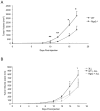
Figure 2. Deletion of Rgs2 in MDSCs retards tumor growth.
(A) Rgs2−/− and C57BL/6 wild type mice were injected with 5×10⁵ 3LL tumor cells subcutaneously in the hindlimb, and tumor size was measured by caliper over time (n = 12 mice per group). This experiment was repeated 3 times. (B) Wild type mice were injected subcutaneously in the hindlimb with 1×10⁵ 3LL cells alone, or 3LL cells combined with 1×10⁴ wild type or Rgs2−/− MDSCs sorted by flow cytometry (>95% purity; data not shown) from spleens of tumor-bearing mice. Tumor growth was measured by caliper over time. n = 8 mice per group. This experiment was performed twice. * p<0.05.
Figure 3
Figure 3. Lack of Rgs2 in endothelial cells does not affect migration, BrdU incorporation, or tube fomation.
Lung microvascular endothelial cells were isolated from lungs of wild type and Rgs2−/− mice and cultured. (A) 1×10⁵ cells were placed in the top chamber of a Transwell, and migrated to growth medium for 4 hours. Cells on the bottom of the Transwell (migrated cells) were fixed, stained with crystal violet, and counted. Experiment was performed 3 times in duplicate. (B) Cells were pulsed with 10 uM BrdU for 2 hours, then collected, processed, and analyzed by flow cytometry. The average percentages of BrdU positive cells was plotted. Experiment was performed 2 times in duplicate. (C) Cells were seeded on top of Matrigel in growth medium and allowed to form tube structures for 48 hours. Branch points were counted, and the average number per field was graphed.
Figure 4
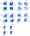
Figure 4. Lack of Rgs2 does not affect populations of mature leukocytes.
Spleens were isolated from non-tumor bearing and 3LL tumor bearing WT and Rgs2−/− mice, processed into single cell suspensions, and labeled with the indicated antibodies, then analyzed by flow cytometry. Experiment was performed 3 times with 3–4 mice per group.
Figure 5
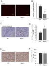
Figure 5. Tumors in Rgs2−/− mice exhibit decreased vascular density and increased cell death.
(A), (C), (E) Sections from size matched 3LL tumors grown in wild type mice and Rgs2 null mice were stained for CD31, active caspase-3 and PCNA, respectively. Representative images are shown. (B), (D), (F) The numbers of CD31 positive vascular structures, active caspase-3 positive cells, and PCNA positive cells, respectively, were quantified in 10 randomly selected fields under microscopy. These experiments were repeated 3–4 times. ** p<0.005, * p<0.05.
Figure 6
Figure 6. Rgs2 deficiency has minimal effects on MDSC expansion and differentiation.
(A) Wild type and Rgs2−/− mice were injected with 1×10⁵ 3LL cells in the hindlimb, and 20 days later, spleens were isolated and analyzed by flow cytometry for Gr-1+CD11b+ MDSCs. This experiment was performed at least 3 times and the graphs shown are results from pooling of 3 mice per group. (B) MDSCs were isolated from spleens of tumor bearing Rgs2−/− and wild type mice using the MACS system, spun onto slides using a cytospin centrifuge, and stained. The slides were read by a hematopathologist in a blind fashion, and cells were categorized by morphology. This experiment was performed 4 times. * p<0.01.
Figure 7
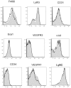
Figure 7. Analysis of cell surface molecules on Gr-1+CD11b+ MDSCs.
Spleens were harvested from 3LL tumor bearing WT or Rgs2−/− mice between days 17–21 post-injection, processed into single cell suspensions, and labeled with antibodies against the indicated cell surface molecules. Representative plots are shown. Experiment was performed 3 times with 3–4 mice per group. Shaded = WT, open = Rgs2−/−.
Figure 8
Figure 8. Rgs2 regulates MCP-1 expression in MDSCs.
MDSCs were isolated from 3LL tumor tissues of wild type or Rgs2−/− mice by magnetic sorting after digestion of the tissues with hyaluronidase and collagenase. (A) Protein lysates from the isolated cells were analyzed using a cytokine array. Each cytokine is detected in duplicate, and intensity was determined using ImageJ software. Positive controls provided on the array were used for normalization. This experiment was performed twice. (B) RNA was extracted from the isolated MDSCs, and analyzed by real time PCR. Beta-actin was used as an internal control. This experiment was repeated 3 times. (C) MDSCs were isolated from tumors of wild type and Rgs2 null mice, and cultured for 48 hours. Culture medium was assayed for MCP-1 protein by ELISA. This experiment was performed in duplicate and repeated 3 times. * p<0.05, ** p<0.00005. (D) MDSCs were isolated from normal spleen and 3LL tumor tissues of wild type mice. Rgs2 and MCP-1 levels were measured by real time PCR. Beta-actin was used as an internal control. This experiment was repeated 2 times. * p<0.05, ** p<0.005.
Figure 9
Figure 9. Angiogenic function of Rgs2 in MDSC is mediated through MCP-1.
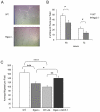
(A) and (B) Wild type and Rgs2−/− MDSCs were isolated from 3LL tumor tissues by magnetic sorting, and incubated overnight at 37°C. 80,000 HUVECs were plated in each well of a 48-well plate on top of Matrigel in the conditioned medium derived from the isolated MDSCs. Representative images are shown at 72 hours, and Vascular network branch points were scored at the times indicated. This experiment was performed in duplicate and repeated twice. (C) MDSCs were isolated from tumors of Rgs2−/− and wild type mice by magnetic sorting, and incubated overnight. Transwells containing 1×10⁵ HUVECs in the top chamber were added and allowed to migrate for 3.5 hours. MCP-1 neutralizing antibody (Ab) (1 ug/ml) was added to wild type cells or 1 ng/ml of recombinant MCP-1 was added to Rgs2−/− cells. This experiment was performed 3 times in duplicate. * p≤0.05, ** p<0.005, *** p<0.001, **** p<0.00005.

References

    1. Tlsty TD, Coussens LM. Tumor stroma and regulation of cancer development. Annu Rev Pathol. 2006;1:119–150. - PubMed
    1. Gabrilovich DI, Nagaraj S. Myeloid-derived suppressor cells as regulators of the immune system. Nat Rev Immunol. 2009;9:162–174. - PMC - PubMed
    1. Sinha P, Clements VK, Bunt SK, Albelda SM, Ostrand-Rosenberg S. Cross-talk between myeloid-derived suppressor cells and macrophages subverts tumor immunity toward a type 2 response. J Immunol. 2007;179:977–983. - PubMed
    1. Yang L, DeBusk LM, Fukuda K, Fingleton B, Green-Jarvis B, et al. Expansion of myeloid immune suppressor Gr+CD11b+ cells in tumor-bearing host directly promotes tumor angiogenesis. Cancer Cell. 2004;6:409–421. - PubMed
    1. Shojaei F, Wu X, Zhong C, Yu L, Liang XH, et al. Bv8 regulates myeloid-cell-dependent tumour angiogenesis. Nature. 2007;450:825–831. - PubMed

Publication types

MeSH terms