. 2011 Apr 11;6(4):e18795.
doi: 10.1371/journal.pone.0018795.

Sequential and coordinated actions of c-Myc and N-Myc control appendicular skeletal development

Affiliations

Sequential and coordinated actions of c-Myc and N-Myc control appendicular skeletal development

Zi-Qiang Zhou et al. PLoS One. .

Abstract

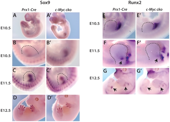
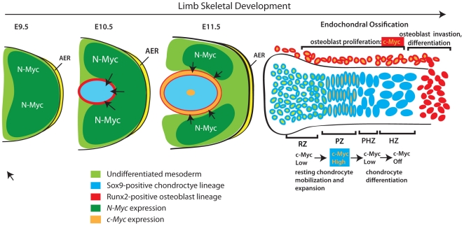
Background: During limb development, chondrocytes and osteoblasts emerge from condensations of limb bud mesenchyme. These cells then proliferate and differentiate in separate but adjacent compartments and function cooperatively to promote bone growth through the process of endochondral ossification. While many aspects of limb skeletal formation are understood, little is known about the mechanisms that link the development of undifferentiated limb bud mesenchyme with formation of the precartilaginous condensation and subsequent proliferative expansion of chondrocyte and osteoblast lineages. The aim of this study was to gain insight into these processes by examining the roles of c-Myc and N-Myc in morphogenesis of the limb skeleton.

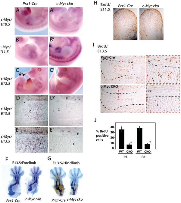
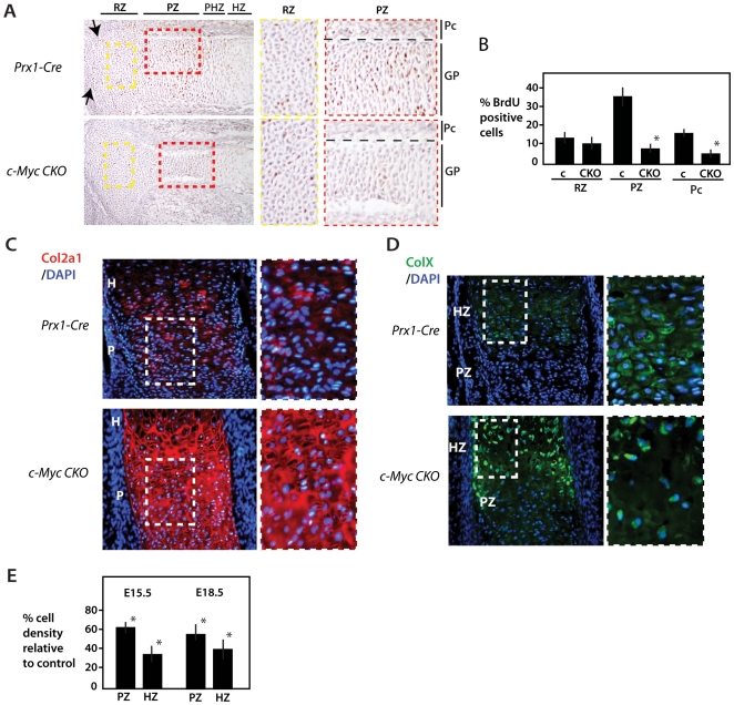
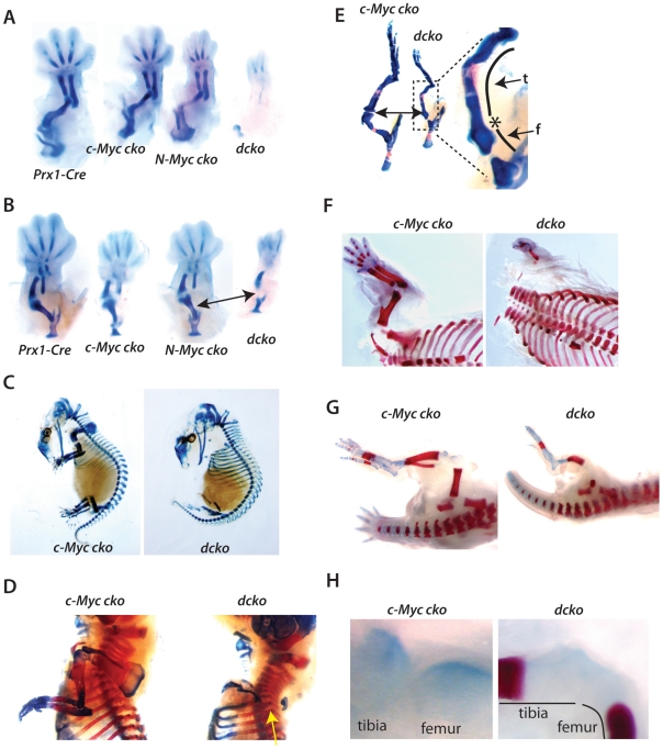
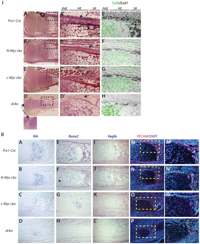
Methodology/principal findings: To investigate c-Myc function in skeletal development, we characterized mice in which floxed c-Myc alleles were deleted in undifferentiated limb bud mesenchyme with Prx1-Cre, in chondro-osteoprogenitors with Sox9-Cre and in osteoblasts with Osx1-Cre. We show that c-Myc promotes the proliferative expansion of both chondrocytes and osteoblasts and as a consequence controls the process of endochondral growth and ossification and determines bone size. The control of proliferation by c-Myc was related to its effects on global gene transcription, as phosphorylation of the C-Terminal Domain (pCTD) of RNA Polymerase II, a marker of general transcription initiation, was tightly coupled to cell proliferation of growth plate chondrocytes where c-Myc is expressed and severely downregulated in the absence of c-Myc. Finally, we show that combined deletion of N-Myc and c-Myc in early limb bud mesenchyme gives rise to a severely hypoplastic limb skeleton that exhibits features characteristic of individual c-Myc and N-Myc mutants.

Conclusions/significance: Our results show that N-Myc and c-Myc act sequentially during limb development to coordinate the expansion of key progenitor populations responsible for forming the limb skeleton.


Conflict of interest statement

Competing Interests: The authors have declared that no competing interests exist.

Figures

Figure 1
Figure 1. c-Myc plays an early role in appendicular skeletal development.
(A–C') Whole mount in situ hybridization of c-Myc in forelimb buds at E10.5, E11.5 and E12.5 of wildtype and Prx1-Cre c-Myc mutant embryos. (D, D') In situ hybridization for c-Myc in sections of proximal tibia at E13.5 of wildtype and Prx1-Cre c-Myc mutant embryos. (E, E') Higher magnification views of proximal tibia sections shown in D and D'. Cartilage and (C) and perichondrium (P) are indicated. (H, I) BrdU staining in limb buds at E11.5 and proximal tibia at E13.5 respectively. (J) Summary of BrdU-positive cells in the proliferative zone (PZ) and perichondrium (Pc).
Figure 2
Figure 2. Limb bud deletion of c-Myc affects Sox9 and Runx2 expression and formation of cartilage anlage.
Analysis of Sox9 expression (A-D') and Runx2 expression (E-G') by in situ hybridization in forelimbs of Prx1-Cre control and c-Myc cko embryos at the indicated stages. Dashed lines delineate the limb bud. (H, H') Alcian blue staining of forelimb and hindlinb cartilage anlage of control and c-Myc cko embryos at E13.5.
Figure 3
Figure 3. Delayed endochondral ossification and suppressed bone growth caused by c-Myc deficiency.
Alcian blue and Alizarin red stained forelimb and hindlimb skeletal preparations at E15.5 (A) and E18.5 (C). Arrows point to primary ossification centers of the humerus and femur to indicate delayed (E15.5) and smaller (E18.5) ossification centers in c-Myc deficient limbs. (B, D) Alcian blue and hemotoxylin and eosin (H&E) stained sections at E15.5 and E16.5. Bars indicate length of primary ossification center. (E) Alizarin red stained tibia at P7 showing small, but fully ossified bone in c-Myc mutants. (F) Measurement of bone length at E18.5. Autopod length was from the tip of digit 3 to the base of the lunate carpal bone.
Figure 4
Figure 4. Control of cell proliferation and cell density in the growth plate by c-Myc.
(A) BrdU labeling of control and Prx1-Cre c-Myc mutant proximal tibia at E16.5. Approximate locations of specific regions of the growth plate (GP) and perichondrium (Pc) are indicated and higher magnification images for the RZ (yellow dashed box) and PZ (red dashed box) are shown at right. The division between cartilage and perichondrium in (A) is marked by the black dashed line. Arrow point to articular regions that show reduced proliferation in c-Myc mutants. (B) Percentage of BrdU labeled cells in comparable regions of the RZ, PZ and perichondrium (Pc) of Prx1-Cre control (c) tibias and Prx1-Cre c-Myc mutant tibias (see Materials and Methods for details). C) Proximal tibias of the indicated stains at E16.5 stained for Col2a1 and DAPI. Higher magnification views are at right. (D) Proximal tibias stained for ColX and DAPI with higher magnification views at right. (E) The percentage change in cell density (cells per unit area) of c-Myc mutants compared to controls in comparable regions of PZ and HZ as determined from sections stained with DAPI alone (see Materials and Methods). *p<0.01.
Figure 5
Figure 5. Disrupted endochondral ossification in c-Myc mutants is linked to decreased numbers of osteoblasts and poor vascularization.
(A) Combined ColXa1 immunohistochemistry and Col1 in situ hybridization at E18.5 showing a smaller hypertrophy zone (marked by ColXa1 – green) and reduced Col1 (dark staining) in the perichondrium of c-Myc mutant tibia. (B) Higher magnification view of the primary ossification center and adjacent perichondrium showing fewer Col1-positive osteoblasts (orange arrow) and a thinner Col1-positive layer in the perichondrium of c-Myc mutant tibia. Red staining is pan-cytokeratin.(C) Runx2 in situ hybridization. (D) Vegfa in situ hybridization showing reduced expression in hypertrophic chondrocytes of c-Myc mutant tibia at E18.5. Approximate locations of proliferative and prehypertrophic zones are indicated and higher magnification images of boxed regions are shown at right. (E) PECAM immunohistochemistry indicating reduced vascularization of the primary ossification center and perichondrium of c-Myc deficient tibia. Red boxed area is shown at higher magnification on right. (F) TUNEL staining for apoptotic cells at the ossification fronts of control and c-Myc mutant tibia.
Figure 6
Figure 6. Decreased expression of key regulators of the transition from proliferation to prehypertrophy in chondrocytes in the absence of c-Myc deletion.
(A–E') In situ hybridization (purple signal) of c-Myc, Fgfr3, IHH, Ptch-1 and PTHrPR in control and c-Myc proximal tibia at E16.5. Arrows point to perichondrial expression of c-Myc, Ptch-1 and PTHrPR expression. Approximate subregions of the growth plate are indicated. (F, G) pRpCTD (pCTD) immunohistochemistry of control and Prx1-Cre c-Myc mutant tibias at E16.5. Higher magnification views are shown at right. Approximate subregions (RZ, PZ and PHZ) of the growth plate are indicated and perichonium (Pc) and growth plate (GP) regions of the higher magnification are indicated.
Figure 7
Figure 7. Combined c-Myc and N-Myc deletion mimics deletion of N-Myc alone in the early limb bud and prevents Sox9 upregulation caused by c-Myc deletion.
(IA–D) BrdU labeling of limb buds at E10.5 for the indicated genotypes. Higher magnification views of boxed areas (red) are shown in A'–D'. Similar results were observed in sections from three different limb buds. (IIA–D') pRpCTD immunohistochemistry of E10.5 limb buds of the indicated genotypes with high (IIIA–D) Comparison of limb size and morphology for the indicated strains at E12.5. (IV) Whole mount in situ hybridization for Fgf8, Shh and Sox9 at E10.5 and Runx2 at E11.5 in Prx1-Cre, c-Myc cko, N-Myc cko and dcko embryos.
Figure 8
Figure 8. Combined c-Myc and N-Myc deletion results in severe limb skeletal agenesis.
(A, B) Alcian blue stained forelimbs respectively of the indicated mouse strains at E13.5. (C) Alcian blue stained c-Myc and dcko mutants at E15.5. (D) Alcian blue and Alizarin red stained preparation of E18.5 embryos showing forelimbs (yellow arrow points to the absence of humerus and distal scapula). (E) E18.5 hindlimb region with higher magnification image at right showing fusion of the femur (f)-tibia (t) joint (*) of dcko embryos. (F) Forelimbs of E20.5 embryos showing absence of humerus and scapula elements in dcko embryos. (G) Hindlimbs of c-Myc and dcko mutants at E20.5. (H) Higher magnification images of the femur-tibia joint region of the E20.5 dcko mutant shown in (G).
Figure 9
Figure 9. Unique and additive contributions of N-Myc and c-Myc to endochondral growth and ossification.
(IA–D') Histological comparison of E18.8 proximal tibia of N-Myc, c-Myc and dcko mutant embryos. Green dashed lines outline the epiphyseal head regions and higher magnification views of the black-boxed regions are shown (A'–D'). Arrow in dcko image (D) indicates fusion between tibia/fibula and femur. (E–H) Combined ColXa1 immunohistochemistry (green) and Col1 in situ hybridization (dark blue) at E18.5. (II) In situ hybridization of Ihh (A–D), Runx2 (E–H) and Vegfa (I–L) and PECAM immunohistochemistry (M–P) in tibia sections at E18.5 from the indicated mouse strains. Yellow boxed regions in M-P are shown in M'–P'.
Figure 10
Figure 10. Model for the combined actions of N-Myc and c-Myc during limb development.
N-Myc expression in the early limb bud promotes expansion of undifferentiated multipotent mesenchymal cells. As the limb bud expands, secreted factors from the ectoderm (e.g. FGF and Wnt family members) that promote N-Myc expression and proliferation in underlying mesenchyme can no longer reach the most central cells and these cells exit the cell cycle and are fated to become Sox9-positive chondro-osteoprogenitors . In addition to cell cycle exit, the hypoxic environment and Hif1α upregulation appears to participate in fate determination steps that initiate the Sox9-positive chondrocyte and Runx2-positive osteoblast lineages. The commencement of low-level c-Myc expression in these lineages is proposed to promote their proliferative expansion, but maintain their multipotent progenitor character. Subsequent elaboration of the growth plate in cartilage anlagen leads to the regional partitioning of c-Myc expression where it acts as part of a program to mobilize resting chondrocytes to proliferate and commit to the chondrocyte terminal differentiation program. Although c-Myc expression in perichondrial osteoblasts likely contributes to their proliferation, by controlling the number of hypertrophic chondrocytes produced, c-Myc impinges on the production of critica l factors produced from these cells that also promotes osteoblast development and endochondral ossification.

Similar articles

Cited by

References

    1. Niswander L. Pattern formation: old models out on a limb. Nat. Rev. . Genet. 2003;4:133–143. - PubMed
    1. Tabin C, Wolpert L. Rethinking the proximodistal axis of the vertebrate limb in the molecular era. Genes Dev. 2007;21:1433–1442. - PubMed
    1. ten Berge D, Brugmann SA, Helms JA, Nusse R. Wnt and FGF signals interact to coordinate growth with cell fate specification during limb development. Development. 2008;135:3247–3257. - PMC - PubMed
    1. Thorogood PV, Hinchliffe JR. An analysis of the condensation process during chondrogenesis in the embryonic chick hind limb. J Embryol Exp Morphol. 1975;33:581–606. - PubMed
    1. Akiyama H, Kim JE, Nakashima K, Balmes G, Iwai N, et al. Osteo-chondroprogenitor cells are derived from Sox9 expressing precursors. Proc Natl Acad Sci U S A. 2005;102:14665–14670. - PMC - PubMed

Publication types

MeSH terms

Substances