

Separase phosphosite mutation leads to genome instability and primordial germ cell depletion during oogenesis
- PMID: 21494564
- PMCID: PMC3073988
- DOI: 10.1371/journal.pone.0018763
Separase phosphosite mutation leads to genome instability and primordial germ cell depletion during oogenesis
Abstract
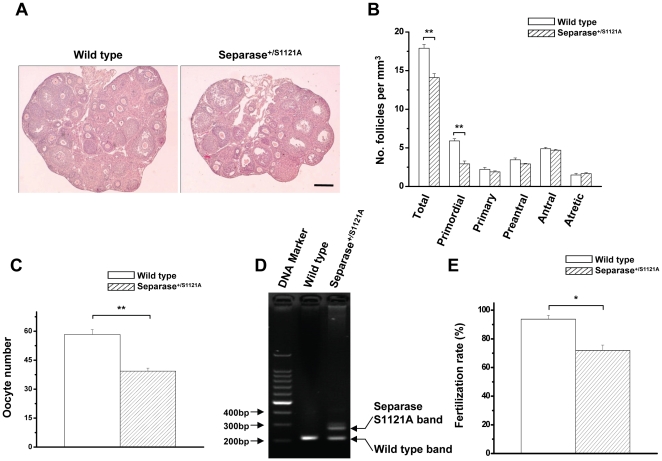
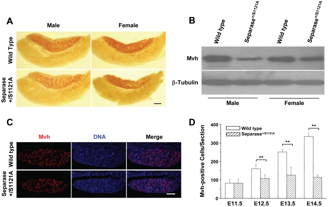
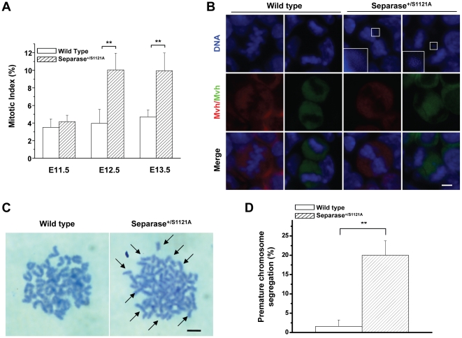
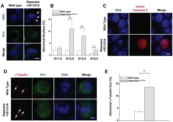
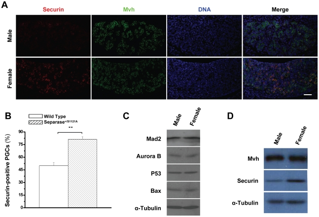
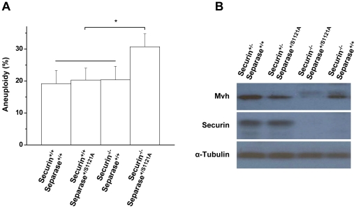
To ensure equal chromosome segregation and the stability of the genome during cell division, Separase is strictly regulated primarily by Securin binding and inhibitory phosphorylation. By generating a mouse model that contained a mutation to the inhibitory phosphosite of Separase, we demonstrated that mice of both sexes are infertile. We showed that Separase deregulation leads to chromosome mis-segregation, genome instability, and eventually apoptosis of primordial germ cells (PGCs) during embryonic oogenesis. Although the PGCs of mutant male mice were completely depleted, a population of PGCs from mutant females survived Separase deregulation. The surviving PGCs completed oogenesis but produced deficient initial follicles. These results indicate a sexual dimorphism effect on PGCs from Separase deregulation, which may be correlated with a gender-specific discrepancy of Securin. Our results reveal that Separase phospho-regulation is critical for genome stability in oogenesis. Furthermore, we provided the first evidence of a pre-zygotic mitotic chromosome segregation error resulting from Separase deregulation, whose sex-specific differences may be a reason for the sexual dimorphism of aneuploidy in gametogenesis.
Conflict of interest statement
Figures
Similar articles
-
Inhibitory phosphorylation of separase is essential for genome stability and viability of murine embryonic germ cells.PLoS Biol. 2008 Jan;6(1):e15. doi: 10.1371/journal.pbio.0060015. PLoS Biol. 2008. PMID: 18232736 Free PMC article.
-
Preimplantation mouse embryos depend on inhibitory phosphorylation of separase to prevent chromosome missegregation.Mol Cell Biol. 2009 Mar;29(6):1498-505. doi: 10.1128/MCB.01778-08. Epub 2009 Jan 5. Mol Cell Biol. 2009. PMID: 19124608 Free PMC article.
-
Securin and separase phosphorylation act redundantly to maintain sister chromatid cohesion in mammalian cells.Mol Biol Cell. 2005 Oct;16(10):4725-32. doi: 10.1091/mbc.e05-03-0190. Epub 2005 Jul 19. Mol Biol Cell. 2005. PMID: 16030258 Free PMC article.
-
[Activity of separase and its regulation].Yi Chuan. 2004 May;26(3):383-6. Yi Chuan. 2004. PMID: 15640025 Review. Chinese.
-
Anaphase topsy-turvy: Cdk1 a securin, separase a CKI.Cell Cycle. 2006 Jan;5(1):11-3. doi: 10.4161/cc.5.1.2296. Epub 2006 Jan 4. Cell Cycle. 2006. PMID: 16340311 Review.
Cited by
-
Kif18a is specifically required for mitotic progression during germ line development.Dev Biol. 2015 Jun 15;402(2):253-262. doi: 10.1016/j.ydbio.2015.03.011. Epub 2015 Mar 28. Dev Biol. 2015. PMID: 25824710 Free PMC article.
-
AIP1-mediated actin disassembly is required for postnatal germ cell migration and spermatogonial stem cell niche establishment.Cell Death Dis. 2015 Jul 16;6(7):e1818. doi: 10.1038/cddis.2015.182. Cell Death Dis. 2015. PMID: 26181199 Free PMC article.
-
Proteolysis in Reproduction: Lessons From Gene-Modified Organism Studies.Front Endocrinol (Lausanne). 2022 May 4;13:876370. doi: 10.3389/fendo.2022.876370. eCollection 2022. Front Endocrinol (Lausanne). 2022. PMID: 35600599 Free PMC article. Review.
-
Structure and Function of the Separase-Securin Complex.Subcell Biochem. 2021;96:217-232. doi: 10.1007/978-3-030-58971-4_4. Subcell Biochem. 2021. PMID: 33252730 Free PMC article. Review.
-
Separase Control and Cohesin Cleavage in Oocytes: Should I Stay or Should I Go?Cells. 2022 Oct 27;11(21):3399. doi: 10.3390/cells11213399. Cells. 2022. PMID: 36359795 Free PMC article. Review.
References
-
- Uhlmann F, Lottspeich F, Nasmyth K. Sister-chromatid separation at anaphase onset is promoted by cleavage of the cohesin subunit Scc1. Nature. 1999;400(6739):37–42. - PubMed
-
- Uhlmann F, Wernic D, Poupart MA, Koonin EV, Nasmyth K. Cleavage of cohesin by the CD clan protease separin triggers anaphase in yeast. Cell. 2000;103(3):375–386. - PubMed
-
- Nasmyth K. Segregating sister genomes: the molecular biology of chromosome separation. Science. 2002;297(5581):559–565. - PubMed
-
- Mailhes JB. Faulty spindle checkpoint and cohesion protein activities predispose oocytes to premature chromosome separation and aneuploidy. Environ Mol Mutagen. 2008;49(8):642–658. - PubMed
-
- Hassold T, Hunt P. To err (meiotically) is human: the genesis of human aneuploidy. Nat Rev Genet. 2001;2(4):280–291. - PubMed
Publication types
MeSH terms
Substances
LinkOut - more resources
Full Text Sources
Molecular Biology Databases
