. 2011 Apr 11;6(4):e18720.
doi: 10.1371/journal.pone.0018720.

CD8+ T cells and IFN-γ mediate the time-dependent accumulation of infected red blood cells in deep organs during experimental cerebral malaria

Affiliations

CD8+ T cells and IFN-γ mediate the time-dependent accumulation of infected red blood cells in deep organs during experimental cerebral malaria

Carla Claser et al. PLoS One. .

Abstract

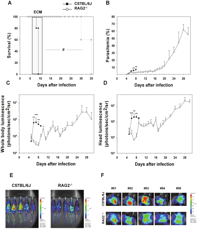
Background: Infection with Plasmodium berghei ANKA (PbA) in susceptible mice induces a syndrome called experimental cerebral malaria (ECM) with severe pathologies occurring in various mouse organs. Immune mediators such as T cells or cytokines have been implicated in the pathogenesis of ECM. Red blood cells infected with PbA parasites have been shown to accumulate in the brain and other tissues during infection. This accumulation is thought to be involved in PbA-induced pathologies, which mechanisms are poorly understood.

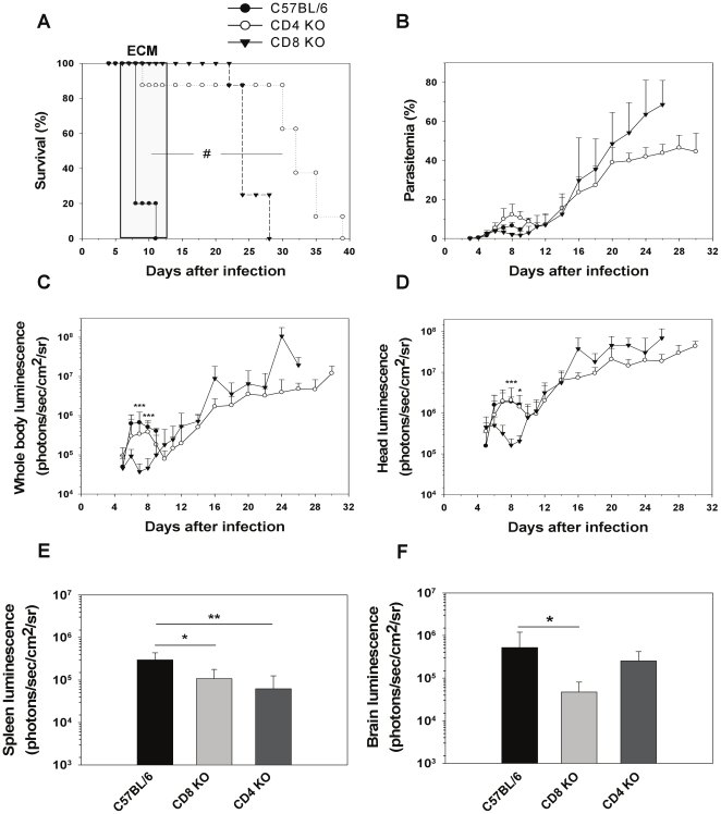
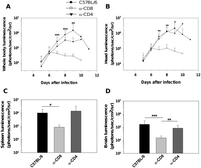
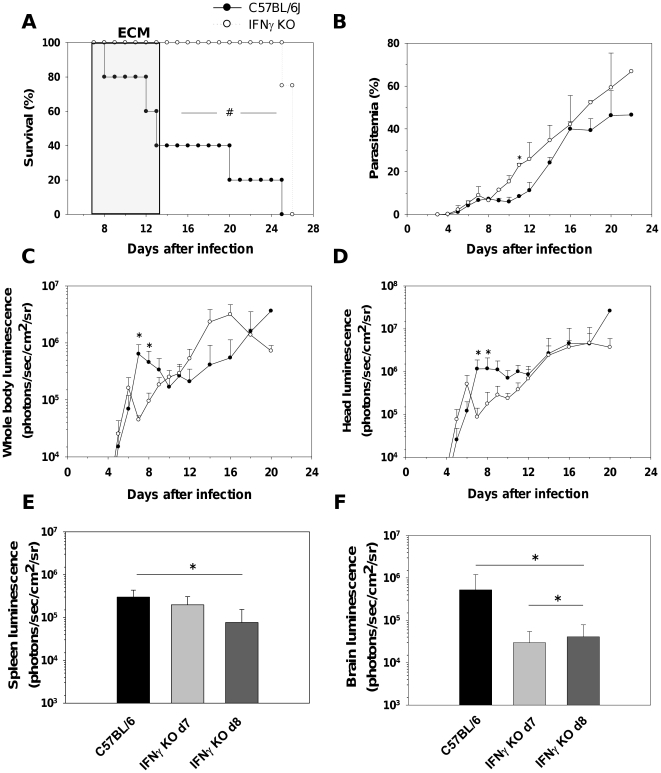
Methods and findings: Using transgenic PbA parasites expressing the luciferase protein, we have assessed by real-time in vivo imaging the dynamic and temporal contribution of different immune factors in infected red blood cell (IRBC) accumulation and distribution in different organs during PbA infection. Using deficient mice or depleting antibodies, we observed that CD8(+) T cells and IFN-γ drive the rapid increase in total parasite biomass and accumulation of IRBC in the brain and in different organs 6-12 days post-infection, at a time when mice develop ECM. Other cells types like CD4(+) T cells, monocytes or neutrophils or cytokines such as IL-12 and TNF-α did not influence the early increase of total parasite biomass and IRBC accumulation in different organs.

Conclusions: CD8(+) T cells and IFN-γ are the major immune mediators controlling the time-dependent accumulation of P. berghei-infected red blood cells in tissues.


Conflict of interest statement

Competing Interests: The authors have read the journal's policy and have the following conflicts: Laurent Rénia is an academic editor of PLoS ONE.

Figures

Figure 1
Figure 1. Decrease in parasite biomass and IRBC accumulation in organs in infected Rag2−/− mice.
A) Survival and (B) parasitemia in WT (n = 5) and Rag2−/− mice (n = 5) infected with PbAluc. Neurologic signs of CM appeared on days 6–12 (shaded area), with death occurring 24–48 h after onset. Parasitemia (%) values are expressed as mean ± SD of 5 mice per group. In vivo bioluminescence imaging quantification of IRBC accumulation in the whole body (C) and head (D) of infected WT and Rag2−/− mice. Mice were injected with luciferin and IRBC accumulation was measured by bioluminescence imaging as described in Materials and Methods. Each bar represents mean luminescence values (log) ± SD of 5 mice. #p<0.01, log-rank test and **p<0.01 and ***p<0.001, Mann Whitney test. Pseudocolor images of whole body (E) and Head (F) from infected WT and RAG2 KO mice.
Figure 2
Figure 2. Decrease in parasite biomass and IRBC accumulation in organs in infected CD8−/− mice.
A) Survival and (B) parasitemia of CD8−/− (n = 8), CD4−/− (n = 8) and WT (n = 10) mice infected with PbAluc. Neurologic signs of CM appeared on days 6–12 (shaded area), with death occurring 24–48 h after onset. Parasitemia (%) values are expressed as mean ± SD. In vivo bioluminescence imaging quantification of IRBC accumulation in the whole body (C) and head (D) of infected mice. Ex vivo quantification by bioluminescence imaging of IRBC accumulation in spleens (E) and brains (F) obtained from five perfused mice per group. Luminescence values (log) as mean ± SD. #p<0.01, log-rank test; *p<0.05, **p<0.01 and ***p<0.001, ANOVA followed by Bonferroni test.
Figure 3
Figure 3. Decrease in parasite biomass and IRBC accumulation in organs from CD8+ T cell-depleted WT mice.
In vivo bioluminescence imaging quantification of IRBC accumulation in the whole body (A) and head (B) of mice infected with PbAluc and depleted of either CD4+ or CD8+ T cells by antibody treatment. Ex vivo quantification by bioluminescence imaging of IRBC accumulation in spleens (C) and brains (D). Data are expressed as mean ± SD. In this experiment, all the control (n = 10) and CD4+-depleted (n = 5) mice infected with PbAluc developed ECM between days 7 and 12, and none of the anti-CD8+-treated (n = 10) infected mice developed CM. On each day that a control-infected mouse developed CM, one CD4 depleted- and one CD8-depleted mouse were sacrificed to determine IRBC accumulation ex vivo. Luminescence values (log) as mean ± SD of 5 mice. *p<0.05, **p<0.01 and ***p<0.001, ANOVA followed by Bonferroni test.
Figure 4
Figure 4. Depletion of myeloid cells does not alter IRBC distribution during ECM.
MAFIA mice were infected with PbAluc and injected on days 5, 6, and 7 with the drug AP20187 as described in Material and Methods. (A) Depletion of granulocytes/monocytes (defined as CD45+CD11b+Gr1+) was assessed by flow cytometry on day 7 post-infection. Data plots presented are from one mouse and similar data were obtained for 4 more mice. (B) Survival and (C) parasitemia of treated and non-treated MAFIA mice (5 per group). Neurologic signs of CM appeared on days 6–12 (shaded area), with death occurring 24–48 h after onset. Parasitemia (%) values are expressed as mean ± SD of 5 mice per group. In vivo bioluminescence imaging quantification of IRBC accumulation in the whole body (D) and head (E) of treated and non-treated infected mice. Luminescence values (log) as mean ± SD of 5 mice.
Figure 5
Figure 5. IFN-γ controls parasite biomass and IRBC accumulation in organs.
(A) Survival and (B) parasitemia of WT and IFN-γ−/− mice (5 mice per group) infected with PbAluc. Parasitemia (%) values are expressed as mean ± SD. In vivo bioluminescence imaging quantification of IRBC accumulation in the whole body (C) and head (D) of WT and IFN-γ −/− mice. Luminescence values (log) as mean ± SD of 5 mice. Ex vivo quantification by bioluminescence imaging of IRBC accumulation in spleens (E) and brains (F) of perfused WT and IFN-γ−/− mice obtained at day 7 and 8 post-infections. Luminescence values (log) as mean ± SD of 5 mice. #p<0.01, log-rank test; *p<0.05, Mann Whitney test; *p<0.05, ANOVA followed by Bonferroni test.

References

    1. Feachem RG, Phillips AA, Hwang J, Cotter C, Wielgosz B, et al. Shrinking the malaria map: progress and prospects. Lancet. 2010;376:1566–1578. - PMC - PubMed
    1. Hay SI, Okiro EA, Gething PW, Patil AP, Tatem AJ, et al. Estimating the global clinical burden of Plasmodium falciparum Malaria in 2007. Plos Med. 2010;7:e1000290. - PMC - PubMed
    1. Severe falciparum malaria. World Health Organization, Communicable Diseases Cluster. Trans R Soc Trop Med Hyg. 2000;94:S1–S90. - PubMed
    1. van der Heyde HC, Nolan J, Combes V, Gramaglia I, Grau GE. A unified hypothesis for the genesis of cerebral malaria: sequestration, inflammation and hemostasis leading to microcirculatory dysfunction. Trends Parasitol. 2006;22:503–508. - PubMed
    1. Idro R, Jenkins NE, Newton CRJC. Pathogenesis, clinical features, and neurological outcome of cerebral malaria. Lancet Neurol. 2005;4:827–840. - PubMed

Publication types

MeSH terms