. 2011 Apr 8;6(4):e18293.
doi: 10.1371/journal.pone.0018293.

A universal system for highly efficient cardiac differentiation of human induced pluripotent stem cells that eliminates interline variability

Affiliations

A universal system for highly efficient cardiac differentiation of human induced pluripotent stem cells that eliminates interline variability

Paul W Burridge et al. PLoS One. .

Abstract

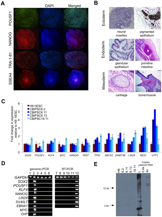
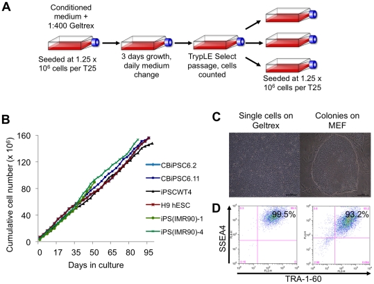
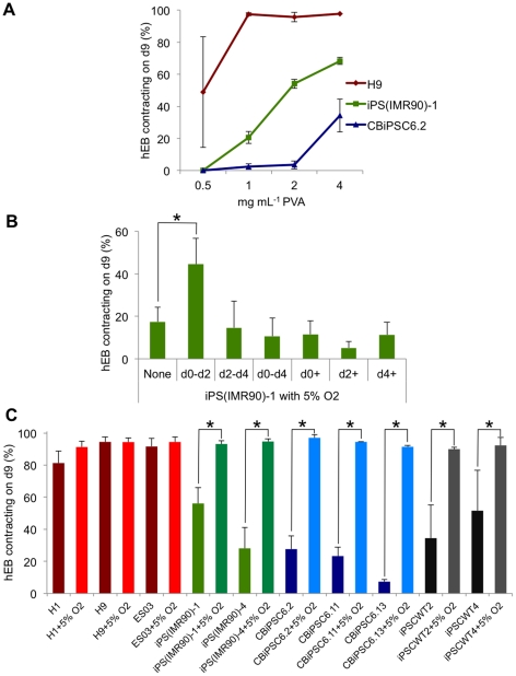
Background: The production of cardiomyocytes from human induced pluripotent stem cells (hiPSC) holds great promise for patient-specific cardiotoxicity drug testing, disease modeling, and cardiac regeneration. However, existing protocols for the differentiation of hiPSC to the cardiac lineage are inefficient and highly variable. We describe a highly efficient system for differentiation of human embryonic stem cells (hESC) and hiPSC to the cardiac lineage. This system eliminated the variability in cardiac differentiation capacity of a variety of human pluripotent stem cells (hPSC), including hiPSC generated from CD34(+) cord blood using non-viral, non-integrating methods.

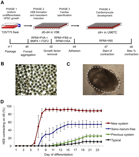
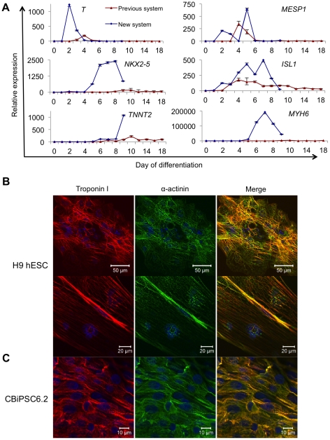
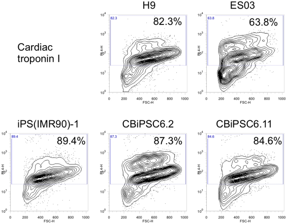
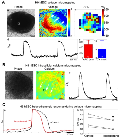
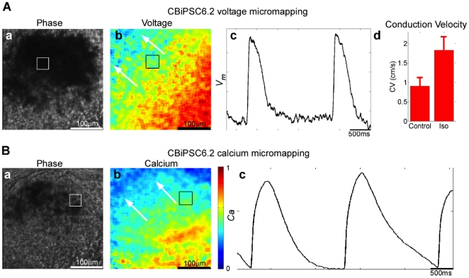
Methodology/principal findings: We systematically and rigorously optimized >45 experimental variables to develop a universal cardiac differentiation system that produced contracting human embryoid bodies (hEB) with an improved efficiency of 94.7±2.4% in an accelerated nine days from four hESC and seven hiPSC lines tested, including hiPSC derived from neonatal CD34(+) cord blood and adult fibroblasts using non-integrating episomal plasmids. This cost-effective differentiation method employed forced aggregation hEB formation in a chemically defined medium, along with staged exposure to physiological (5%) oxygen, and optimized concentrations of mesodermal morphogens BMP4 and FGF2, polyvinyl alcohol, serum, and insulin. The contracting hEB derived using these methods were composed of high percentages (64-89%) of cardiac troponin I(+) cells that displayed ultrastructural properties of functional cardiomyocytes and uniform electrophysiological profiles responsive to cardioactive drugs.

Conclusion/significance: This efficient and cost-effective universal system for cardiac differentiation of hiPSC allows a potentially unlimited production of functional cardiomyocytes suitable for application to hPSC-based drug development, cardiac disease modeling, and the future generation of clinically-safe nonviral human cardiac cells for regenerative medicine.


Conflict of interest statement

Competing Interests: The authors have declared that no competing interests exist.

Figures

Figure 1
Figure 1. Systematic optimization of cardiac differentiation of human pluripotent stem cells.
(A) Schematic of the optimized cardiac differentiation system demonstrating: Phase 1, uniform growth of hESC/hiPSC as monolayers. Phase 2 (d0–d2), aggregation of 5000 single cell hESC or hiPSC in chemically defined RPMI+PVA medium in V96 plates. Phase 3 (d2–d4), cardiac specification using FBS or hSA containing medium. Phase 4 (d4+), cardiac development, hEB are allowed to adhere to U96 tissue culture treated plates in RPMI+INS. (B) Typical d2 hEB formed using the forced aggregation procedure in RPMI-PVA to demonstrate homogeneity in hEB size. Scale bar = 500 µm. (C) Typical d9 contracting hEB formed using the optimized cardiac differentiation method with the contracting area circled, note minimal fibroblast outgrowth. Scale bar = 200 µm. (D) Efficiency of generation of contracting hEB produced in this system (New system, n = 48), with comparisons to xeno- and serum-free conditions (Xeno-/serum-free, n = 5), our previous method (Previous system, n = 7), and typically used methods (Typical, n = 9). Error bars, ± S.E.M.
Figure 2
Figure 2. Generation of non-viral hiPSC from CD34+ cord blood progenitors.
(A) Representative immunocytochemistry of pluripotency markers POU5F1 (OCT4), NANOG, TRA-1-81, and SSEA4 in hiPSC line CBiPSC6.2 after >20 passages. (B) Representative hematoxylin and eosin staining of teratoma sections derived from CBiPSC6.2 after >20 passages demonstrating ectodermal, endodermal and mesodermal lineage differentiation. All CBiPSC clones in these studies formed similar teratomas. (C) Real-time RT-PCR studies p15 CBiPSC lines for endogenous pluripotency genes using primers that distinguish endogenous expression from transgenes (see Methods). (D) The presence of plasmid transgene sequences examined by PCR at p11 in CBiPSC6.2, CBiPSC6.11, CBiPSC6.13, CBiPSC19.11 (lanes 3–6, respectively) and negative control H9 hESC (p48) (lane 1) compared to positive control early cultures from p2 (lane 2). RT-PCR analysis of selected plasmid sequences in p11 CBiPSC6.2, CBiPSC6.11, CBiPSC6.13, CBiPSC19.11 (lanes 8–11, respectively) and negative control H9 hESC (lane 7) and p2 cultures (lane 12). (E) Genomic Southern blot analysis for episomal vector backbone integration in lines CBiPSC6.2, CBiPSC6.11, CBiPSC6.13, CBiPSC19.11 (p15) (lanes 2–5, respectively), H9 hESC (p55) (lane 1). Combination 6 episomal vector DNA was diluted as positive control to the equivalents of 0.4 and 4 integrations per haploid genome (0.4× and 4×). L: 1 kb plus ladder. These studies were also conducted for non-viral adult fibroblast-derived hiPSC lines iPSCWT2 and iPSCWT4 with similar results (Machairaki et al., in preparation).
Figure 3
Figure 3. Controlled growth of hPSC lines for reproducible cardiac differentiation.
(A) Schematic of monolayer hESC/hiPSC culture technique. This monolayer technique uses conditioned medium prepared in a defined manner, single-cell passaging, automated cell counting, plating cells at a known density, and passaging every three days. (B) Stable growth rates of H9 hESC, viral fetal fibroblast-derived hiPSC lines iPS(IMR90)-1 and iPS(IMR90)-4, non-viral CD34+ cord blood-derived hiPSC lines CBiPSC6.2, CBiPSC6.11, and non-viral adult fibroblast-derived hiPSC line iPSCWT4. (C) The homogenous culture phenotype seen when culturing H9 hESC as feeder-free monolayers (left), compared in comparison to typical colony morphology of cells grown in co-culture with MEF (right). (D) Comparable SSEA4 and TRA-1-60 expression of H9 hESC growing as monolayer cultures (left) or colonies grown in co-culture with MEF (right).
Figure 4
Figure 4. PVA supplementation and staged exposure to physiological oxygen eliminates interline variability of cardiac differentiation.
(A) Increasing the concentration of PVA from 1 mg mL−1 to 4 mg mL−1 PVA in the d0–d2 media formulation enhanced the differentiation of viral fibroblast-derived hiPSC line iPS(IMR90)-1, and non-viral cord blood-derived hiPSC line CB-iPSC6.2, whilst not affecting H9 hESC (n = 3). (B) Exposure of iPS(IMR90)-1 hEB to physiological (5%) oxygen tensions from differentiation d0–d2 also enhanced cardiac differentiation (n = 3, p<0.005). Identical conditions did not improve already highly efficient H9 hESC differentiation. (C) Combining both physiological oxygen tension and 4 mg mL−1 PVA between d0–d2 eliminated interline differentiation variability in hiPSC derived using both viral- and non-viral-techniques (n≥3, p<0.005). Error bars, ± S.E.M.
Figure 5
Figure 5. Characterization of hPSC cardiomyocyte differentiation.
(A) Comparison of real-time RT-PCR for markers of mesoderm (T, MESP1), cardiac progenitors (NKX2.5, ISL1), and cardiomyocytes (TNNT2, MYH6) during hESC differentiation using either the Previous system or New system. Analysis was performed using the ΔΔCt method with relative expression calculated using d0 of differentiation (hESC samples) as baseline. 18S RNA expression was used for normalization. Primers are shown in Table S1. (B) Immunocytochemistry for cardiac markers in hEB differentiated from H9 hESC. Troponin I (red), α-actinin (green) and DAPI (blue) at low power (top panels) demonstrating unaligned striations throughout the hEB and higher power (lower panels) demonstrating area of aligned striation. (C) Immunocytochemistry for cardiac markers in hEB differentiated from CBiPSC6.2.
Figure 6
Figure 6. Intracytoplasmic flow cytometry analysis of cardiomyocyte marker expression.
Expression of cardiac troponin I in contracting hEB from differentiated H9 hESC, ES03 hESC, viral fibroblast-derived line iPS(IMR90)-1 hiPSC, and non-viral CD34+ cord blood-derived hiPSC lines CBiPSC6.2 and CBiPSC6.11. For each sample the entire U-well contents of one 96-well plate was enzymatically digested into a single cell suspension and analyzed by flow cytometry.
Figure 7
Figure 7. Electrophysiological characterization H9 hESC contracting hEB using optical mapping.
(A) Voltage micromapping. a, Phase contrast image of H9 hEB at 4× magnification. b, Voltage activation map (arrows indicate direction of electrical wave propagating across hEB). c, Action potential duration (APD) map. d, Representative transmembrane potential (Vm) trace at position denoted by the small square in a and b during 0.5 Hz field stimulation. e, Mean APD and conduction velocity (CV) measurements from 19 hEB (error bars represent ± s.d.). Coefficient of variation (COV, population s.d. divided by mean) for APD was 0.30 and for CV was 0.88 across hEB population. COV within an individual hEB was calculated from multiple APD measurements across all of the recording sites for that hEB (panel A, c) and was 0.042±0.030 (s.d.) when averaged across 19 hEB. (B) Intracellular calcium micromapping. a, Phase map of hEB at 6× magnification. b, Calcium map (arrows indicate direction of propagating calcium wave). c, Representative intracellular calcium (Cai) trace at position denoted by the box in a and b. (C) The beta-adrenergic agonist isoproterenol shortened the mean APD in all 4 hEB by an average of 23±8 ms (mean ± s.d.). * indicates p≈0.01 in a paired Student's t-test.
Figure 8
Figure 8. Electrophysiological characterization non-viral cord blood-derived hiPSC contracting hEB differentiated using optical mapping.
(A) Voltage micromapping. Phase image of CBiPSC6.2 hEB at 4× magnification. b. Voltage map, hEB were stained with di-4-ANEPPS (a voltage-sensitive fluorescent dye), and electrically field-stimulated at the same pacing rate (arrows indicate direction of electrical wave propagating across hEB). c. Action potentials during 0.5 Hz field stimulation. d. Average conduction velocity (CV) measurements (mean ± standard deviation) for control and 15 min after addition of 50 µM isoproterenol during 0.5 Hz field stimulation. (B) Calcium micromapping. a. Phase image of beating hEB. b. Calcium map (arrows indicate direction of calcium wave propagating across beating hEB). c. Representative calcium transient (Ca) waveforms during 0.5 Hz field stimulation. Box in a and b denote site of recording in c.

Comment in

References

    1. Hansson EM, Lindsay ME, Chien KR. Regeneration next: toward heart stem cell therapeutics. Cell Stem Cell. 2009;5:364–377. - PubMed
    1. Kehat I, Gepstein A, Spira A, Itskovitz-Eldor J, Gepstein L. High-resolution electrophysiological assessment of human embryonic stem cell-derived cardiomyocytes: a novel in vitro model for the study of conduction. Circ Res. 2002;91:659–661. - PubMed
    1. Xu C, Police S, Rao N, Carpenter MK. Characterization and enrichment of cardiomyocytes derived from human embryonic stem cells. Circ Res. 2002;91:501–508. - PubMed
    1. Mummery C, Ward-van Oostwaard D, Doevendans P, Spijker R, van den Brink S, et al. Differentiation of human embryonic stem cells to cardiomyocytes: role of coculture with visceral endoderm-like cells. Circulation. 2003;107:2733–2740. - PubMed
    1. Braam SR, Tertoolen L, van de Stolpe A, Meyer T, Passier R, et al. Prediction of drug-induced cardiotoxicity using human embryonic stem cell-derived cardiomyocytes. Stem Cell Res. 2010;4:107–116. - PubMed

Publication types

MeSH terms