. 2011 Apr 8;6(4):e18633.
doi: 10.1371/journal.pone.0018633.

Ibudilast, a pharmacologic phosphodiesterase inhibitor, prevents human immunodeficiency virus-1 Tat-mediated activation of microglial cells

Affiliations

Ibudilast, a pharmacologic phosphodiesterase inhibitor, prevents human immunodeficiency virus-1 Tat-mediated activation of microglial cells

Michelle Kiebala et al. PLoS One. .

Abstract

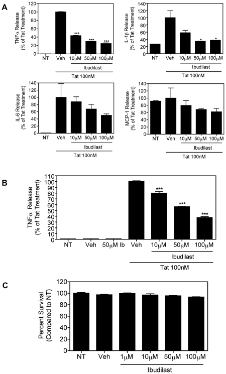
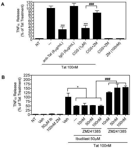
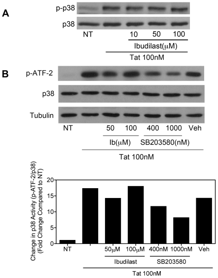
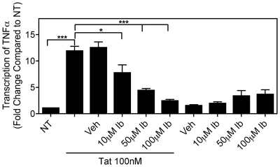
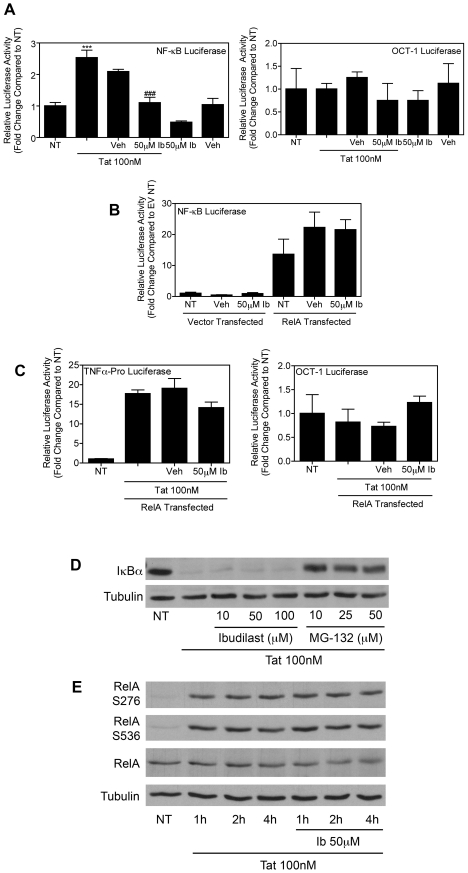
Human Immunodeficiency Virus-1 (HIV-1)-associated neurocognitive disorders (HAND) occur, in part, due to the inflammatory response to viral proteins, such as the HIV-1 transactivator of transcription (Tat), in the central nervous system (CNS). Given the need for novel adjunctive therapies for HAND, we hypothesized that ibudilast would inhibit Tat-induced excess production of pro-inflammatory cytokines, such as tumor necrosis factor-alpha (TNFα) in microglial cells. Ibudilast is a non-selective cyclic AMP phosphodiesterase inhibitor that has recently shown promise as a treatment for neuropathic pain via its ability to attenuate glial cell activation. Accordingly, here we demonstrate that pre-treatment of both human and mouse microglial cells with increasing doses of ibudilast inhibited Tat-induced synthesis of TNFα by microglial cells in a manner dependent on serine/threonine protein phosphatase activity. Ibudilast had no effect on Tat-induced p38 MAP kinase activation, and blockade of adenosine A(2A) receptor activation did not reverse ibudilast's inhibition of Tat-induced TNFα production. Interestingly, ibudilast reduced Tat-mediated transcription of TNFα, via modulation of nuclear factor-kappa B (NF-κB) signaling, as shown by transcriptional activity of NF-κB and analysis of inhibitor of kappa B alpha (IκBα) stability. Together, our findings shed light on the mechanism of ibudilast's inhibition of Tat-induced TNFα production in microglial cells and may implicate ibudilast as a potential novel adjunctive therapy for the management of HAND.


Conflict of interest statement

Competing Interests: The authors have declared that no competing interests exist.

Figures

Figure 1
Figure 1. Ibudilast dose-dependently inhibits Tat-induced pro-inflammatory cytokine production in microglial cells.
A, Human microglial cells (1×10⁵) were left untreated (NT) or were treated with Tat (100 nM) for 8 h with or without pre-treatment for 30 min. with increasing concentrations of ibudilast or vehicle (Veh), as indicated. TNFα, IL-1β, IL-6, and MCP-1 levels in culture supernatants were analyzed by Multi-Plex cytokine array, as described in Methods. The Tat+Veh-treated samples were set to 100% and all other samples were compared to this value. Results are shown as mean ± SD of values derived from two replicates from a single representative experiment. Statistical significance (***, p<0.001; *, p<0.05) is indicated, as compared to Tat+Veh-treated cells. The average cytokine/chemokine concentration of the Tat+Veh-treated sample was as follows; TNFα: 8166 pg/mL, IL-1β: 3.3 pg/mL, IL-6: 6027 pg/mL, and MCP-1: 1480 pg/mL. B, Similarly, murine microglial cells (BV-2; 1.2×10⁵) were left untreated (NT) or were treated with Tat (100 nM) for 8 h with or without pre-treatment for 30 min. with increasing concentrations of ibudilast or vehicle (Veh), as indicated. TNFα release was measured by ELISA. Results are shown as mean ± SEM of values derived from three replicates from a single representative experiment; three total experiments were performed. Statistical significance (p<0.001) is indicated, as compared to Tat+Veh-treated cells (***). The TNFα concentration of the Tat+Veh-treated sample was 1861 pg/mL. C, BV-2 cells (1.2×10⁵) were left untreated (NT) or were treated with increasing concentrations of ibudilast or vehicle (Veh), as indicated, for 8 h. The MTT assay was used as a measure of cell viability. Percent survival was calculated as compared to the untreated sample. Results are shown as mean ± SEM of values derived from four replicates from a single representative experiment; two total experiments were performed.
Figure 2
Figure 2. Inhibition of Tat-induced TNFα production by ibudilast is not reversed by blockade of adenosine A2A receptor.
A, BV-2 cells (1.5×10⁵) were left untreated (NT) or were treated with Tat (100 nM) for 8 h with or without pre-treatment for 30 min. with either anti-Tat or control IgG antibodies (8 µg/mL) or adenosine A2A receptor agonist, CGS21680 (CGS, 1 µM) or antagonist, ZM241385 (ZM, 100 nM), as indicated. TNFα release was measured by ELISA. The Tat-treated samples were set to 100% and all other samples were compared to this value (the TNFα concentration for this sample was 3475 pg/µg total protein content). Results are shown as mean ± SEM of values derived from four replicates from a single representative experiment; two total experiments were performed. Statistical significance (p<0.001) is indicated, as compared to Tat-treated cells (***) or as compared to Tat+CGS-treated cells (###). B, BV-2 cells (1.2×10⁵) were left untreated (NT) or were treated with Tat (100 nM) for 8 h with or without pre-treatment for 30 min. with ibudilast (Ib, 50 µM) or vehicle (Veh) alone or together with increasing concentrations of the adenosine A2A receptor antagonist, ZM241385, as indicated. TNFα release was measured by ELISA. The Tat+Veh-treated samples were set to 100% and all other samples were compared to this value (the TNFα concentration for this sample was 696.2 pg/mL). Results are shown as mean ± SEM of values derived from three replicates from a single representative experiment; two total experiments were performed. Statistical significance is indicated, as compared to Tat+Veh-treated cells (p<0.05, *) or as compared to Tat+ZM241385-treated cells (p<0.001, ###).
Figure 3
Figure 3. Ibudilast does not inhibit Tat-induced activation of p38 MAP kinase.
A, BV-2 cells (1.2×10⁵) were left untreated (NT) or were treated with Tat (100 nM) for 15 min. with or without pre-treatment for 5 min. with increasing concentrations of ibudilast, as indicated. Whole cell lysates were subjected to immunoblot analysis using either phospho-p38 MAPK-specific (upper panel) or p38 MAPK-specific (lower panel) antibodies. The results of a single representative experiment are shown; three total experiments were performed. B, BV-2 cells (2.4×10⁵) were left untreated (NT) or were treated with Tat (100 nM) for 20 min. with or without pre-treatment for 10 min. with increasing concentrations of ibudilast or the p38 MAP kinase inhibitor, SB203580, as indicated. Whole cell lysates were subjected to immunoprecipitation with an immobilized phospho-p38 MAP kinase antibody. Precipitated p38 was incubated with an ATF-2 fusion protein substrate. Phosphorylated ATF-2 levels were determined by immunoblot analysis with a phospho-ATF-2-specific antibody (top panel). Whole cell lysate supernatants collected after immunoprecipitation were subjected to immunoblot analysis using p38-specific (middle panel) or α-Tubulin-specific (lower panel) antibodies. Protein levels were quantified using ImageJ software (bottom graph). Phospho-ATF-2 levels were normalized to total p38 MAPK levels and fold change compared to NT was calculated. The results of a single representative experiment are shown; two total experiments were performed.
Figure 4
Figure 4. Ibudilast inhibits Tat-induced TNFα production in a protein phosphatase dependent manner.
A, BV-2 cells (1.2×10⁵) were left untreated (NT) or were treated with Tat (100 nM) for 8 h with or without pre-treatment for 30 min. with ibudilast (Ib, 50 µM) or vehicle (Veh) alone or together with the protein phosphatase inhibitor, okadaic acid (Oka, 50 nM). TNFα release was measured by ELISA. The Tat+Veh-treated samples were set to 100% and all other samples were compared to this value (the TNFα concentration for this sample was 1861 pg/mL). Results are shown as mean ± SEM of values derived from three replicates from a single representative experiment; two total experiments were performed. Statistical significance is indicated, as compared to Tat+Veh-treated cells (p<0.001, ***) or as compared to Tat+Ib-treated cells (p<0.01, ##). B, Serine/Threonine phosphatase activity was measured in whole cell lysates from BV-2 cells (4.6×10⁵) treated with ibudilast (50 µM) for the indicated periods of time. Results are shown as mean ± SEM of values derived from three replicates from a single representative experiment; two total experiments were performed. Statistical significance (p<0.001) is indicated, ***. C, BV-2 cells (1.2×10⁵) were left untreated (NT) or were treated with ibudilast (50 µM) for 8 h or 24 h, as indicated. Whole cell lysates were subjected to immunoblot analysis using either PP2Ac-specific (upper panel) or α-Tubulin-specific (lower panel) antibodies. The results of a single representative experiment are shown; two total experiments were performed.
Figure 5
Figure 5. Ibudilast inhibits Tat-induced TNFα transcript levels.
BV-2 cells (2.5×10⁵) were left untreated (NT) or were treated with Tat (100 nM) for 4 h with or without pre-treatment for 30 min. with increasing concentrations of ibudilast (Ib) or vehicle (Veh). Total RNA was collected, reverse transcribed using oligo-dT primers, and subjected to Real-Time SYBR Green RT-PCR amplification. Fold induction of TNFα mRNA species was normalized to those of GAPDH and presented as a function of the expression level in NT samples. Data represent mean ± SEM of values derived from three replicates from a single representative experiment. Statistical significance (***, p<0.001 or *, p<0.05) is denoted as compared to Tat-treated samples.
Figure 6
Figure 6. Ibudilast inhibits Tat-induced TNFα production via modulation of NF-κB signaling.
A, BV-2 cells (10×106) were transiently transfected using Nucleofector (Amaxa/Lonza) with an NF-κB-dependent luciferase reporter plasmid. 16 h post-transfection, cells were left untreated or were treated with Tat (100 nM) for 8 h with or without pre-treatment for 30 min. with 50 µM ibudilast (Ib) or vehicle (Veh). Luciferase activity in whole cell lysates was determined. Results are shown as mean ± SEM of values derived from three replicates from a single representative experiment; two total experiments were performed. Statistical significance is denoted (***, p<0.001) as compared to NT samples or (###, p<0.001) as compared to Tat-treated samples. A parallel transfection with a luciferase reporter containing responsive elements of the OCT-1 transcription factor upstream of a firefly luciferase gene was included as a control for the specificity of ibudilast's inhibition of NF-κB transcriptional activity. B, BV-2 cells (10×106) were transiently transfected using Nucleofector (Amaxa/Lonza) with an NF-κB-dependent luciferase reporter plasmid alone or together with either a RelA-encoding plasmid or a vector control plasmid. 16 h post-transfection, cells were left untreated or were treated with 50 µM ibudilast (Ib) or vehicle (Veh) for 8 h. Luciferase activity in whole cell lysates was determined. Results are shown as mean ± SEM of values derived from three replicates from a single representative experiment; two total experiments were performed. Co-transfection with a Renilla luciferase reporter plasmid was included as a control for transfection efficiency. For each sample the firefly luciferase reading was divided by the Renilla luciferase reading. C, BV-2 cells (10×106) were transiently transfected using Nucleofector (Amaxa/Lonza) with a plasmid containing a luciferase reporter gene under transcriptional control of the mouse TNFα promoter region, alone or together with a RelA-encoding plasmid. 16 h post-transfection, cells were left untreated or were treated with Tat (100 nM) for 8 h with or without pre-treatment for 30 min. with 50 µM ibudilast (Ib) or vehicle (Veh). Luciferase activity in whole cell lysates was determined. Results are shown as mean ± SEM of values derived from three replicates from a single representative experiment; two total experiments were performed. A parallel transfection with the OCT-1 luciferase reporter was included as a control for the specificity of ibudilast's inhibition of TNFα promoter activation. D, BV-2 cells (1.2×10⁵) were left untreated (NT) or were treated with Tat (100 nM) for 15 min. with or without pre-treatment for 30 min. with increasing concentrations of ibudilast or MG-132, as indicated. Whole cell lysates were subjected to immunoblot analysis using either IκBα-specific (upper panel) or α-Tubulin-specific (lower panel) antibodies. The results of a single representative experiment are shown; three total experiments were performed. E, BV-2 cells (1.2×10⁵) were left untreated (NT) or were treated with Tat (100 nM) for the indicated periods of time with or without pre-treatment for 30 min. with increasing concentrations of ibudilast, as indicated. Whole cell lysates were subjected to immunoblot analysis using RelA phospho-serine 276-specific (first panel), RelA phospho-serine 536-specific (second panel), total RelA-specific (third panel), or α-Tubulin-specific (fourth panel) antibodies. The results of a single representative experiment are shown; two total experiments were performed.

Similar articles

Cited by

References

    1. Resnick L, Berger JR, Shapshak P, Tourtellotte WW. Early penetration of the blood-brain-barrier by HIV. Neurology. 1988;38:9–14. - PubMed
    1. Kaul M, Lipton SA. Mechanisms of neuronal injury and death in HIV-1 associated dementia. Curr HIV Res. 2006;4:307–318. - PubMed
    1. Nath A, Sacktor N. Influence of highly active antiretroviral therapy on persistence of HIV in the central nervous system. Curr Opin Neurol. 2006;19:358–361. - PubMed
    1. Tozzi V, Balestra P, Bellagamba R, Corpolongo A, Salvatori MF, et al. Persistence of neuropsychologic deficits despite long-term highly active antiretroviral therapy in patients with HIV-related neurocognitive impairment: prevalence and risk factors. J Acquir Immune Defic Syndr. 2007;45:174–182. - PubMed
    1. McArthur JC. HIV dementia: an evolving disease. J Neuroimmunol. 2004;157:3–10. - PubMed

Publication types

MeSH terms