. 2011 Apr 8;6(4):e18674.
doi: 10.1371/journal.pone.0018674.

Methylation and loss of Secreted Frizzled-Related Protein 3 enhances melanoma cell migration and invasion

Affiliations

Methylation and loss of Secreted Frizzled-Related Protein 3 enhances melanoma cell migration and invasion

Elin J Ekström et al. PLoS One. .

Abstract

Background: Wnt signaling is important in development and can also contribute to the initiation and progression of cancer. The Secreted Frizzled Related Proteins (SFRPs) constitute a family of Wnt modulators, crucial for controlling Wnt signaling. Here we investigate the expression and role of SFRP3 in melanoma.

Methodology/principal findings: We show that SFRP3 mRNA is down-regulated in malignant melanoma tumors as compared to normal/benign tissue. Furthermore, we found that SFRP3 expression was lost in the malignant melanoma cell lines, A2058, HTB63 and A375, but not in the non-transformed melanocyte cell line, Hermes 3A. Methylated CpG rich areas were detected in the SFRP3 gene in melanoma cell lines and their SFRP3 expression could be restored using the demethylating agent, 5'aza-deoxycytidine. Addition of recombinant SFRP3 to melanoma cells had no effect on viable cell numbers, but decreased cell migration and invasion. Wnt5a signaling has been shown to increase the migration and invasion of malignant melanoma cells, and high expression of Wnt5a in melanoma tumors has been connected to a poor prognosis. We found that recombinant SFRP3 could inhibit Wnt5a signaling, and that it inhibited melanoma cell migration and invasion in a Wnt5a-dependent manner.

Conclusion/significance: We conclude that SFRP3 functions as a melanoma migration and invasion suppressor by interfering with Wnt5a signaling.


Conflict of interest statement

Competing Interests: TA and VS are shareholders of WntResearch. This does not alter the authors' adherence to all the PLoS ONE policies on sharing data and materials.

Figures

Figure 1
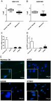
Figure 1. SFRP3 mRNA and protein levels are reduced in melanoma.
A, Microarray data from Gene Expression Omnibus (GEO) dataset GDS1375, Normal/benign vs. Melanoma. B, Microarray data from GEO dataset GDS1989, Normal/benign vs. Melanoma. Bars represent the 95th and 5th percentile. C, Basal SFRP3 mRNA expression in cell lines. A human non-transformed immortalized melanocyte cell line, Hermes 3A, and three malignant melanoma cell lines: A375, A2058 and HTB63 cells were analyzed for their SFRP3 mRNA expression using RT-QPCR. The data is representative of 3 independent experiments and the error bars represent standard deviation (SD) within one experiment. D, Basal SFRP3 protein levels in media from the indicated cell lines. The SFRP3 protein levels were normalized against total protein content. Error bars represent Standard Error of the Mean (SEM) (n = 4). Statistical analysis determined the following p-values; Hermes 3A vs. A375 p = 0,0025, Hermes 3A vs. A2058 p = 0,0051 and Hermes 3A vs HTB63 p = 0,1091. E, Basal SFRP3 protein expression in cell lines. Melanocytes and melanoma cells were analyzed for SFRP3 expression using immunofluorescence. Representative immunofluorescence images from 3 independent experiments. * = p<0.05, **** = p<0.0001.
Figure 2
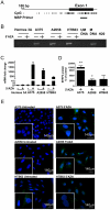
Figure 2. Methylation of SFRP3 in melanoma cell lines.
A, Schematic representation of the promoter region together with the first exon of the SFRP3 gene. The CpG rich areas and the site of the methylation specific PCR primer are indicated. B, Methylation specific PCR. Hermes 3A, A375, A2058 and HTB63 cells were treated for 72 h with 5 µM 5′AZA and analyzed using methylation specific PCR. Universally unmethylated DNA (UM DNA) and methylated DNA (M DNA) were used as negative and positive controls, respectively. The image is representative of at least 3 independent experiments. C, SFRP3 mRNA expression after demethylation treatment. Hermes 3A, A375, A2058 and HTB63 cells were treated with 5′AZA as in B and relative mRNA expression of SFRP3 was analyzed using RT-QPCR. The graph is representative of data from 3 independent experiments and error bars denote SD within one experiment. The relative expression of each cell line is normalized to the untreated control of the same cell line. D, SFRP3 protein levels in the media from the indicated cell lines after demethylation treatment. A375, A2058 and HTB63 cells were treated as described for B and the SFRP3 protein content analyzed using the ELISA kit as described in Figure 1. The results are given as a percentage compared to non-demethylated control cells. Error bars represent SEM (n = 4). E, SFRP3 protein expression in the melanoma cell lines. The indicated melanoma cell lines were analyzed for their SFRP3 content by fluorescence microscopy after demethylation treatment using non-treated cells as controls. Representative immunofluorescence images from at least 3 independent experiments. * = p<0.05, ** = p<0.01.
Figure 3
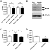
Figure 3. Recombinant SFRP3 decreases migration and invasion of melanoma cells.
A, Invasion assay using Matrigel invasion chambers. A2058 cells were resuspended in serum free media and treated in the upper chamber with 1 µg/ml of rSFRP3 or carrier (0.1% BSA in PBS) and allowed to invade for 24 h. The error bars represent SEM, (n = 5). B, Wound healing assay. A2058 cells were treated with either 1 µg/ml of rSFRP3 (dashed line, squares) or carrier (full line, circles). Pictures were taken of the scratches at 0, 16, 24 and 48 h. Data is given as percentage of wound area closed at each time point. The error bars represent SEM, (n = 5). C, Migration assay, A2058 cells were treated with either 1 µg/ml of rSFRP3 or carrier in serum-free media and migration of non-dividing individual cells was analyzed manually by tracking the speed of cells for 12 h. The error bars represent SEM, (n = 5). * = p<0.05. D, Wnt5a protein expression analysis. Wnt5a expression was analyzed in A2058 cells and HTB63 cells using western blotting.
Figure 4
Figure 4. SFRP3 can inhibit Wnt5a signaling.
A, Topflash reporter assay. A2058 cells transiently transfected with Topflash and Fopflash plasmids. Where appropriate, cells were pre-treated with 1 µg/ml rSFRP3 or carrier, and after 24 h cells were treated with the indicated combinations of Wnt ligands (0.2 µg/ml rWnt5a and 0.05 µg/ml rWnt3a) as indicated for an additional 24 h. The image is representative of 5 independent experiments and the error bars represent SD within one experiment. B, Measurement of intracellular Ca2+ signaling. rWnt5a (0.1 µg/ml, addition indicated by arrows) was added to A2058 cells pre-treated with carrier (upper panel) or 1 µg/ml rSFRP3 (lower panel). Traces are representative images of 4 separate experiments. C, ΔCa2+ ratio values from A2058 cells pre-treated with either carrier or 1 µg/ml rSFRP3 and then stimulated with 0.1 µg/ml rWnt5a. Error bars represent SD (n = 4). ** = p<0,01.
Figure 5
Figure 5. SFRP3 inhibits migration in melanoma cells by antagonizing Wnt5a.
A, Migration assay using Wnt5a knockdown cells. siRNA transfected A2058 cells were grown in serum-free media for 24 h and then treated with 1 µg/ml rSFRP3 or carrier. Migration was analyzed using time-lapse microscopy for 24 h. The error bars represent SD, (n = 4). B, Wnt5a knockdown. The knockdown of Wnt5a in A2058 cells were analyzed using western blot but also by RT-PCR (Figure S4). C, Migration assay using rWnt5a and rSFRP3 in A2058 cells. Cells were pre-treated with either carrier or 1 µg/ml rSFRP3 in serum-free media for 24 h. The cells were then treated with rWnt5a 0.2 µg/ml and analyzed using time-lapse microscopy as in A for 24 h. The error bars represent SD, (n = 4). D, SFRP3 conditioned medium and migration assay. HTB63 cells were transfected with pCMV6-Empty-Vector or pCMV6-SFRP3 and kept in serum-free media for 24 h. These media were transferred to non-transfected HTB63 cells that were then analyzed using time-lapse microscopy as in A. Error bars represent SD (n = 3). * = p<0.05.

Similar articles

Cited by

References

    1. Chien AJ, Conrad WH, Moon RT. A Wnt survival guide: from flies to human disease. J Invest Dermatol. 2009;129:1614–1627. - PMC - PubMed
    1. Mikels A, Minami Y, Nusse R. Ror2 receptor requires tyrosine kinase activity to mediate Wnt5A signaling. J Biol Chem. 2009;284:30167–30176. - PMC - PubMed
    1. O'Connell MP, Fiori JL, Xu M, Carter AD, Frank BP, et al. The orphan tyrosine kinase receptor, ROR2, mediates Wnt5A signaling in metastatic melanoma. Oncogene. 2009;29:34–44. - PMC - PubMed
    1. O'Connell MP, Weeraratna AT. Hear the Wnt Ror: how melanoma cells adjust to changes in Wnt. Pigment Cell Melanoma Res. 2009;22:724–739. - PMC - PubMed
    1. Rao TP, Kuhl M. An updated overview on Wnt signaling pathways: a prelude for more. Circ Res. 2010;106:1798–1806. - PubMed

Publication types