. 2011 Apr 8;6(4):e18689.
doi: 10.1371/journal.pone.0018689.

The critical role of IL-34 in osteoclastogenesis

Affiliations

The critical role of IL-34 in osteoclastogenesis

Zhi Chen et al. PLoS One. .

Abstract

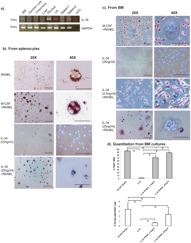
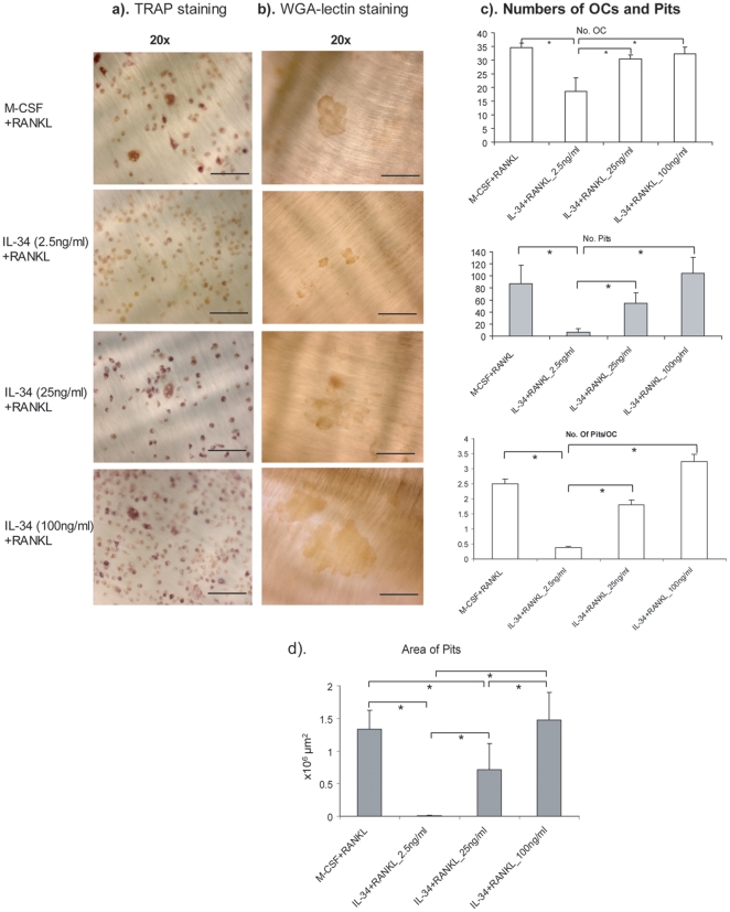
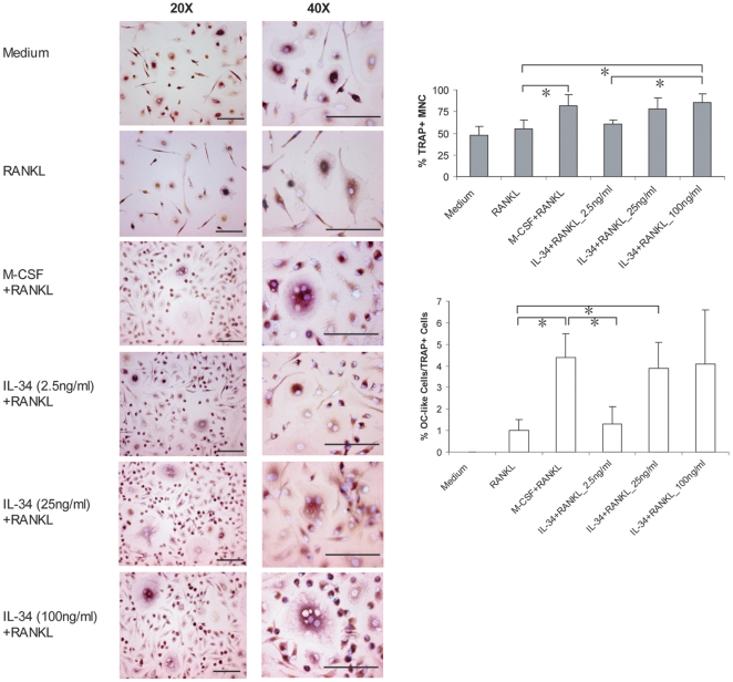
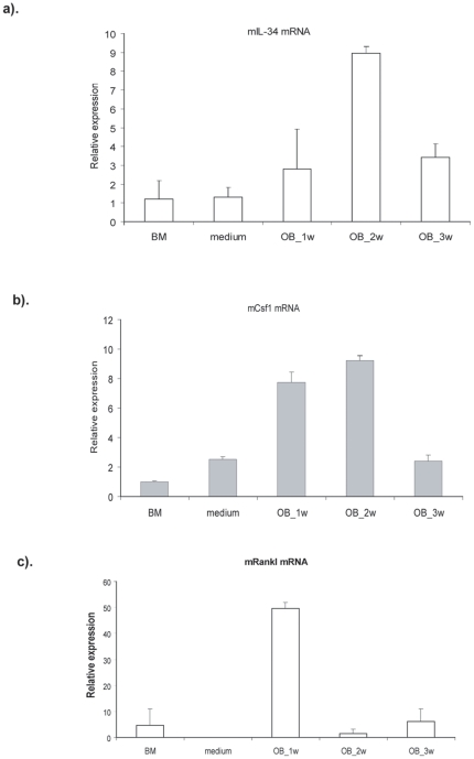
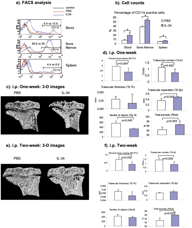
It has been widely believed that the cytokines required for osteoclast formation are M-CSF (also known as CSF-1) and RANKL. Recently, a novel cytokine, designated IL-34, has been identified as another ligand of CSF1R. This study was to explore the biological function, specifically osteoclastogenesis and bone metabolism, of the new cytokine. We produced recombinant mouse IL-34 and found that together with RANKL it induces the formation of osteoclasts both from splenocytes as well as dose-dependently from bone marrow cells in mouse and these cells also revealed bone resorption activity. It also promotes osteoclast differentiation from human peripheral blood mononucleated cells. Finally, we show that systemic administration of IL-34 to mice increases the proportion of CD11b+ cells and reduces trabecular bone mass. Our data indicate that IL-34 is another important player in osteoclastogenesis and thus may have a role in bone diseases. Strategies of targeting CSF1/CSF1R have been developed and some of them are already in preclinical and clinical studies for treatment of inflammatory diseases. Our results strongly suggest the need to revisit these strategies as they may provide a new potential pharmaceutical target for the regulation of bone metabolism in addition to their role in the treatment of inflammatory diseases.


Conflict of interest statement

Competing Interests: The authors have declared that no competing interests exist.

Figures

Figure 1
Figure 1. IL-34 combined with RANKL promotes the differentiation of mouse osteoclast-like cells from splenocytes and bone marrow.
(a). The expression of Il34 in mouse tissues was performed by RT-PCR. Total RNA from mouse tissues were isolated. BM: bone marrow. LN: lymph nodes. NTC: no template control. (b). Splenocytes were isolated from 6–8-week-old Balb/c mice and cultured for 9 days in the presence of RANKL (100 ng/ml) alone, IL-34 (25 ng/ml) alone or RANKL combined either with 25 ng/ml of M-CSF or with 25 ng/ml of IL-34. The cells were fixed with 3% paraformaldehyde and were subjected to TRAP and Hoechst 33258 staining. (c). Bone marrow cells were isolated from the femurs and tibias of 6–8-week-old Balb/c mice. After depletion of adherent stromal cells by culturing the cells overnight with α-MEM media, the nonadherent bone marrow cells were cultured for 9 days in the presence of 25 ng/ml of IL-34 alone, 25 ng/mL recombinant M-CSF or with a different concentration of rmIL-34 (2.5 ng/ml, 25 ng/ml) and 100 ng/ml of RANKL. Three independent experiments were performed. All images in this study were acquired by Leica DMRB microscope and Leica DC300F digital camera system. Representative images are shown with a magnification of 20× or 40×. Bars, 100 µm. (d). The number of TRAP+ mononuclear cells and TRAP+ multinucleared cells (≥3 nuclei, shown as OC-like cells) were counted under microscopy. Data shown were average number counted from four wells. One-way ANOVA analysis was performed and was followed by Turkey's and Dunnett's post-hoc test by using SPSS statistic analysis software. *: The mean difference is significant at the 0.05 level.
Figure 2
Figure 2. In vitro differentiated osteoclasts by IL-34 and RANKL show dose-dependent bone resorption activity.
Mouse nonadherent bone marrow cells were cultured on bone slices for 9 days in the presence of RANKL and M-CSF or RANKL with 2.5 ng/ml, 25 ng/ml, 100 ng/ml of IL-34. The bone slices with cells were fixed and stained for TRAP, and all TRAP-positive multinucleated cells were counted and analyzed under a microscope. The cells were removed followed by WGA-lectin staining for pits. Subsequent counting of resorption pits was performed with a microscope. Representative images of TRAP staining (a) and WGA-lectin staining (b) under different culturing conditions. Representative images are shown with a maginification of 20×. Bars, 100 µm. (c). Histogram of number of osteoclast-like cells, number of pits under different culturing conditions and number of pits/osteoclast (n = 5). (d). Area of Pits was quantitated using an Olympus microscope connected to a computer and the OsteoMeasure program (version 3.21; OsteoMetrics, Atlanta, GA, USA), n = 5. One-way ANOVA analysis was performed and was followed by Turkey's and Dunnett's post-hoc test by using SPSS statistic analysis software. *: The mean difference is significant at the 0.05 level.
Figure 3
Figure 3. In vitro differentiation of human osteoclasts by IL-34 and RANKL.
CD14+ human mononuclear cells were isolated from human peripheral blood followed by purification with anti-CD14 coated meganetic beads. The cells were cultured with RANKL alone or RANKL combined either with M-CSF or with human IL-34 at the indicated concentrations for 9 days. Cells were fixed followed by TRAP and Hoechst 33258 staining. Three independent experiments were performed. Representative images are shown with a maginification of 20× and 40×. Bars, 100 µm. The right panel showed the quantitation of the number of TRAP+ mononuclear cells and TRAP+ multinucleared cells (≥3 nuclei, shown as OC-like cells) counted under microscopy. Data shown were average number counted from four wells. One-way ANOVA analysis was performed and was followed by Turkey's and Dunnett's post-hoc test by using SPSS statistic analysis software. *: The mean difference is significant at the 0.05 level.
Figure 4
Figure 4. IL-34 is expressed by osteoblasts.
Bone marrow cells were cultured in phenol red-free α-MEM media supplemented with 10% fetal calf serum, l0 nmol/L dexamethasone, 50 µg/mL ascorbic acid, and 10 mmol/L sodium β-glycerophosphate. Cells were harvested after 1, 2 and 3 weeks culture and total RNA was isolated. The expression of Rankl, Csf1 and Il34 were detected by real-time quantitative RT-PCR. Hprt was used as an endogenous control. Three independent experiments were performed.
Figure 5
Figure 5. Systemic administration of IL-34 to mice increases the number of monocytes/macrophages and reduces bone mass in vivo.
rmIL-34 was injected into 8-week old Balb/c mice daily at 250 ug/kg, i.p. After one week or two weeks of injections, the mice were sacrificed. Cells from the peripheral blood, bone marrow and spleen were treated with ACK buffer to lyse red blood cells, followed by anti-CD11b-PE staining. CD11b-positive cells were detected by FACSCalibur. (a). Representative FACS histogram images from CD11b-PE staining from peripheral blood, bone marrow cells and splenocytes. (b). Cell numbers of CD11b-positive cells in peripheral blood, bone marrow and spleen from PBS or IL-34 injected mice (n = 4). * indicating p<0.05. (c). Representative 3-D micro-CT images of the metaphyseal region of proximal tibias from mice injected with PBS or IL-34 for one week. (d). Histograms of 3-D trabecular structure parameters from micro-CT analysis (n = 3). (e). Representative 3-D micro-CT images of the metaphyseal region of proximal tibias from mice injected with PBS or IL-34 for two weeks. (f). Histograms of 3-D trabecular structure parameters from micro-CT analysis (n = 4).

References

    1. Lacey DL, Timms E, Tan HL, Kelley MJ, Dunstan CR, et al. Osteoprotegerin ligand is a cytokine that regulates osteoclast differentiation and activation. Cell. 1998;93(2):165–176. - PubMed
    1. Teitelbaum SL. Bone resorption by osteoclasts. Science. 2000;289(5484):1504–1508. - PubMed
    1. Roodman GD. Regulation of osteoclast differentiation. Ann N Y Acad Sci. 2006;1068:100–109. - PubMed
    1. Kong YY, Yoshida H, Sarosi I, Tan HL, Timms E, et al. OPGL is a key regulator of osteoclastogenesis, lymphocyte development and lymph-node organogenesis. Nature. 1999;397(6717):315–323. - PubMed
    1. Pearse RN, Sordillo EM, Yaccoby S, Wong BR, Liau DF, et al. Multiple myeloma disrupts the TRANCE/ osteoprotegerin cytokine axis to trigger bone destruction and promote tumor progression. Proc Natl Acad Sci U S A. 2001;98(20):11581–11586. - PMC - PubMed

Publication types

MeSH terms