. 2011 Apr 6;6(4):e18562.
doi: 10.1371/journal.pone.0018562.

Wnt/β-catenin signaling enhances cyclooxygenase-2 (COX2) transcriptional activity in gastric cancer cells

Affiliations

Wnt/β-catenin signaling enhances cyclooxygenase-2 (COX2) transcriptional activity in gastric cancer cells

Felipe Nuñez et al. PLoS One. .

Abstract

Background: Increased expression of the cyclooxygenase-2 enzyme (COX2) is one of the main characteristics of gastric cancer (GC), which is a leading cause of death in the world, particularly in Asia and South America. Although the Wnt/β-catenin signaling pathway has been involved in the transcriptional activation of the COX2 gene, the precise mechanism modulating this response is still unknown.

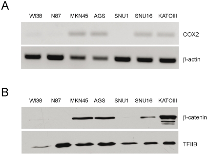
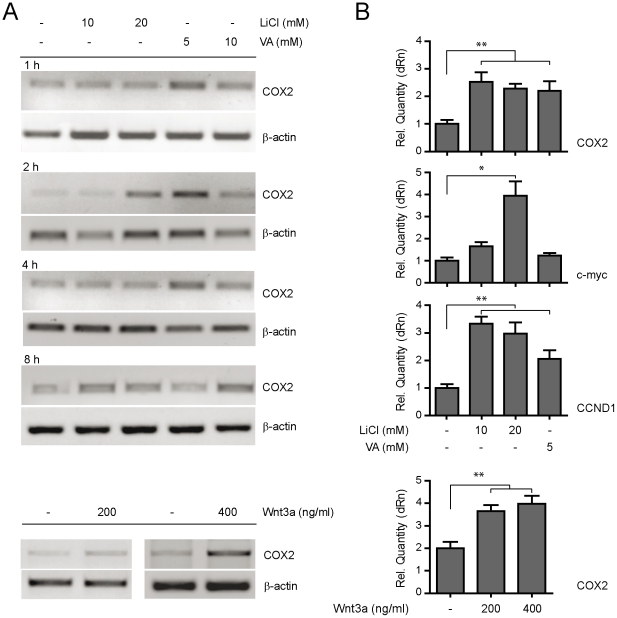
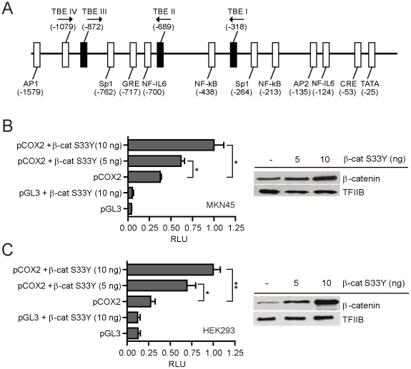
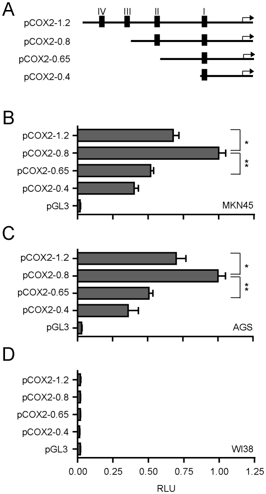
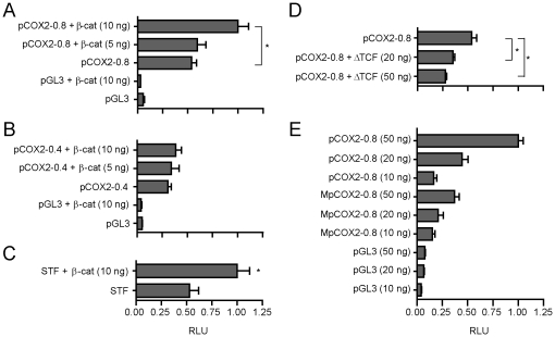
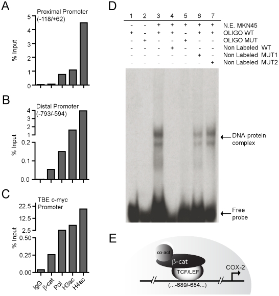
Methodology/principal findings: Here we studied the transcriptional regulation of the COX2 gene in GC cell lines and assessed whether this phenomenon is modulated by Wnt/β-catenin signaling. We first examined the expression of COX2 mRNA in GC cells and found that there is a differential expression pattern consistent with high levels of nuclear-localized β-catenin. Pharmacological treatment with either lithium or valproic acid and molecular induction with purified canonical Wnt3a significantly enhanced COX2 mRNA expression in a dose- and time-dependent manner. Serial deletion of a 1.6 Kbp COX2 promoter fragment and gain- or loss-of-function experiments allowed us to identify a minimal Wnt/β-catenin responsive region consisting of 0.8 Kbp of the COX2 promoter (pCOX2-0.8), which showed maximal response in gene-reporter assays. The activity of this pCOX2-0.8 promoter region was further confirmed by site-directed mutagenesis and DNA-protein binding assays.

Conclusions/significance: We conclude that the pCOX2-0.8 minimal promoter contains a novel functional T-cell factor/lymphoid enhancer factor (TCF/LEF)-response element (TBE Site II; -689/-684) that responds directly to enhanced Wnt/β-catenin signaling and which may be important for the onset/progression of GC.


Conflict of interest statement

Competing Interests: The authors have declared that no competing interests exist.

Figures

Figure 1
Figure 1. COX2 gene expression and nuclear localization of β-catenin in GC cells.
(A) COX2 mRNA expression in control (WI38) and GC cell-lines (MKN45, AGS, SNU1, SNU16, KATOIII and N87). Total RNA was extracted from cultured cells and semiquantitative RT-PCR was used to determine COX2 and β-actin RNA levels as an internal control. Twenty-six cycles were chosen as an adequate PCR cycle. (B) Nuclear levels of β-catenin protein in the same cell-lines, as shown in (A), were examined through Western Blot analysis using nuclear extracts. The TFIIB general transcription factor was used as an internal control.
Figure 2
Figure 2. Pharmacological and molecular enhancement of COX2 expression via Wnt/β-catenin signaling.
(A) COX2 mRNA expression levels in MKN45 cells treated for 1 to 8 h with either 10–20 mM of lithium (LiCl) or 5–10 mM of Valproic acid (VA) were evaluated through RT-PCR. As an internal control, β-actin levels were determined. (B) Q-PCR for COX2, c-myc and CCND1 mRNA expression stimulated pharmacologically with lithium (LiCl) and valproate (VA) during 2 h (top panel). Bottom panel, Q-PCR for COX2 in MKN45 cells stimulated with purified Wnt3a for 2 h. Q-PCR results were expressed as Relative Quantity (dRn) and β-actin mRNA levels were used as the internal control. Each figure corresponds to at least 3 independent experiments. Statistical significance was determined through ANOVA test (* p<0.05, ** p<0.01).
Figure 3
Figure 3. Human pCOX2-1.6 promoter activity in response to Wnt/β-catenin signaling.
(A) Schematic drawing depicting the genomic context of ca. 1.6 Kbp from the transcriptional start site (TSS) of the human COX2 promoter, including the location of known transcriptional regulators (white boxes) and the position of the three novel TCF/LEF-binding elements (TBE: core CTTTG; black boxes) determined in silico. (B & C) Gene reporter assays in MKN45 (B) and HEK293 (C) cells co-transfected with 10 ng of pCOX2 and increasing concentrations of a constitutively active β-catenin (S33Y) protein (left panel). Cells were co-transfected with 1 ng of PRL-SV40 Renilla as an internal control. Promoter activity was normalized as the ratio between firefly luciferase and Renilla luciferase units. RLU: Relative Luciferase Units. Each figure corresponds to at least three independent experiments. Statistical significance was determined through ANOVA test (* p<0.05, ** p<0.01). Nuclear levels of β-catenin protein were examined in same cell lines through Western Blot analysis (right panel). The TFIIB general transcription factor was used as an internal control.
Figure 4
Figure 4. pCOX2-0.8 as a minimal COX2 promoter with maximum basal response in GC cells.
(A) Schematic representation of pCOX2 deletions. (B–D) Gene reporter assay in MKN45 (B), AGS (C) and WI38 (D) cell lines transiently transfected with 50 ng pCOX deletions (pCOX2-1.2; pCOX2-0.8; pCOX2-0.65 and pCOX2-0.4) and 50 ng of empty vector. In all experiments cells were transfected with 1 ng of PRL-SV40 Renilla as an internal control. Promoter activity was normalized as the ratio between firefly luciferase and Renilla luciferase units. RLU: Relative Luciferase Units. Each figure corresponds to at least three independent experiments. Statistical significance was determined through ANOVA test (* p<0.05, ** p<0.01).
Figure 5
Figure 5. Wnt/β-catenin signaling modulates pCOX2-0.8 activity in MKN45 cells.
Gene reporter assays in MKN45 cells transiently transfected with either 10 ng pCOX2-0.8 (A) or pCOX2-0.4 (B), plus 5–10 ng of β-catenin S33Y and 10 ng of empty vector as control. (C) SuperTOPFlash (STF; 10 ng) was co-transfected with 10 ng of β-catenin S33Y as a positive control for Wnt/β-catenin signaling activity. (D) Effect of a dominant negative TCF-4 (ΔTCF) construct on the activity of pCOX2-0.8. MKN45 cells were transiently co-transfected with 50 ng pCOX2-0.8 and 20–50 ng of ΔTCF. (E) Comparison between the activity of the wild-type pCOX2-0.8 and a mutant MpCOX2-0.8 construct, containing mutated residues in the TBE Site II in the pCOX2-0.8 promoter as background. Cells were transfected with increasing concentrations of pCOX2-0.8, MpCOX-08, or equal amounts of empty vector as a control. In all experiments 1 ng of PRL-SV40 Renilla was transfected as an internal control. Promoter activity was normalized as the ratio between firefly luciferase and Renilla luciferase units. RLU: Relative Luciferase Units. Each figure corresponds to at least three independent experiments. Statistical significance was determined through ANOVA test (* p<0.05, ** p<0.01).
Figure 6
Figure 6. Binding of β-catenin to the TBE Site II (−689/−684) in the COX2 promoter.
(A–C) ChIP assays in MKN45 cells using specific antibodies for β-catenin (β-cat), polymerase II (Pol), H3 and H4 acetylated histones (H3ac; H4ac) and immunoglobulin G (IgG). Quantification was done by real time PCR using specific primers for the proximal promoter (PP) region (A), the TBE Site II (−689/−684) in the human COX2 promoter (B) and a TBE site within the c-myc promoter, as a positive control (C). (D) EMSA assay in nuclear extracts from MKN45 cells. DNA-protein complexes formed by incubating nuclear extracts (N.E.) from MKN45 cells with radiolabelled probes containing the intact and a mutated TBE Site II (lanes 3 and 5) were resolved in native polyacrylamide gels at 5% and revealed through autoradiography. To determine the specificity of the binding a 50 times excess of non-radiolabelled wild-type (line 4) and mutant (6 and 7) oligonucleotides were added. Lanes 1 and 2 correspond to the wild-type and mutant radiolabelled oligonucleotides without incubation with N.E. These tests are representative of three independent experiments. E. Model depicting the molecular mechanism by which Wnt/β-catenin signaling may contribute to the expression of the human COX2 gene.

Similar articles

Cited by

References

    1. Crew KD, Neugut AI. Epidemiology of gastric cancer. World J Gastroenterol. 2006;12:354–362. - PMC - PubMed
    1. Parkin DM. International variation. Oncogene. 2004;23:6329–6340. - PubMed
    1. Ushijima T, Sasako M. Focus on gastric cancer. Cancer Cell. 2004;5:121–125. - PubMed
    1. Konturek PC, Konturek SJ, Brzozowski T. Gastric cancer and Helicobacter pylori infection. J Physiol Pharmacol. 2006;57(Suppl 3):51–65. - PubMed
    1. Lee JH, Abraham SC, Kim HS, Nam JH, Choi C, et al. Inverse relationship between APC gene mutation in gastric adenomas and development of adenocarcinoma. Am J Pathol. 2002;161:611–618. - PMC - PubMed

Publication types