

Spatial differences in the presence of FOXP3+ and GranzymeB+ T cells between the intra- and extravascular compartments in renal allograft vasculopathy
- PMID: 21494640
- PMCID: PMC3071842
- DOI: 10.1371/journal.pone.0018656
Spatial differences in the presence of FOXP3+ and GranzymeB+ T cells between the intra- and extravascular compartments in renal allograft vasculopathy
Abstract
Background: Allograft vasculopathy (AV) and native atherosclerosis (NA) share the presence of a T-cell mediated inflammatory response, but differ in overall plaque morphology and growth rate. We studied the distribution and frequency of regulatory- and cytotoxic T cells in the arterial intima lesions in both conditions.
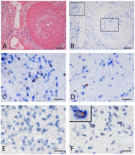
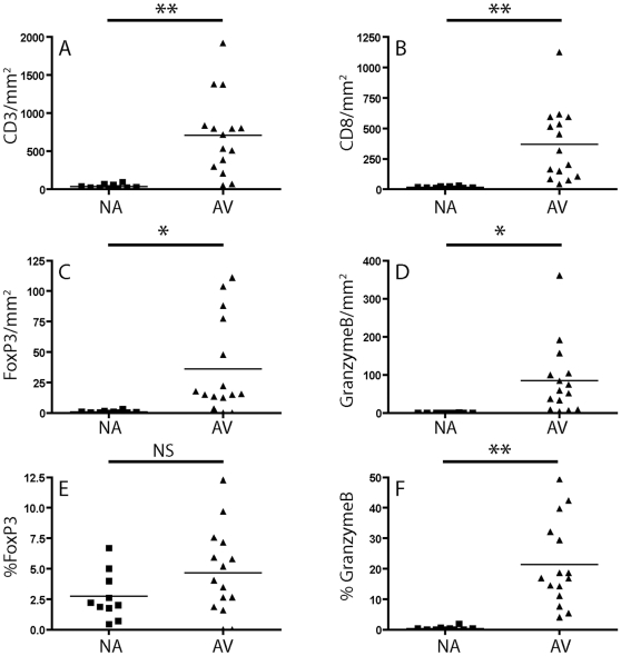
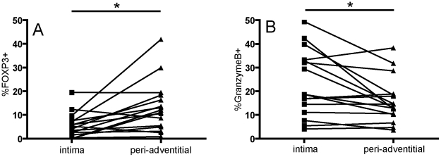
Methodology/principal findings: The study is based on vessels of 15 explanted human renal allografts with AV and 10 carotid artery plaques obtained at surgery. Distribution and frequency of cytotoxic- and regulatory T cells, as identified by the expression of Granzyme B (GrB) and FOXP3 was established in NA and AV. Furthermore, we compared the distribution of these cells in AV with the perivascular, interstitial renal tissue using immunohistochemistry. The total number of T cells was much higher in AV than in NA lesions (711±135 and 37±8 CD3/mm(2) respectively, p<0.005, mean, ± SEM). Total numbers of FOXP3(+) regulatory cells were also significantly increased in AV (36±10 and 0.9±0.3 FOXP3(+)/mm(2) p<0.05), but relative numbers, expressed as a percentage of the total number of CD3(+) T cells ((FOXP3(+)/CD3(+)) ×100), were not significantly different (4.6%±0.9 and 2.7%±0.6). GrB(+) cells were rare in NA, but significantly increased numbers of GrB(+) cells were found in AV lesions (85±24 and 0.2±0.1 GrB(+)/mm(2), p<0.05). Perivascular tissues in the allografts showed a higher relative frequency of FOXP3(+) cells than adjacent intimal lesions (14.0%±2.7 and 4.6%±0.9, respectively, p<0.05), but a lower frequency of GrB(+) cytotoxic T cells (16.1%±2.7 and 22.6%±3.6, p<0.05).
Conclusions: Similar to NA, AV is characterized by a low frequency of intimal FOXP3(+) regulatory T cells. Moreover, significant spatial differences exist in the distribution of functional T cell subsets between the intra- and extravascular micro-environments of the graft.
Conflict of interest statement
Figures
References
-
- Paul LC. Chronic rejection of organ allografts: magnitude of the problem. Transplantation Proceedings. 1993;25:2024–2025. - PubMed
-
- Sharples LD, Caine N, Mullins P, Scott JP, Solis E, et al. Risk factor analysis for the major hazards following heart transplantation–rejection, infection, and coronary occlusive disease. Transplantation. 1991;52:244–252. - PubMed
-
- Rahmani M, Cruz RP, Granville DJ, McManus BM. Allograft vasculopathy versus atherosclerosis. Circulation Research. 2006;99:801–815. - PubMed
-
- de Boer OJ, Becker AE, van der Wal AC. T lymphocytes in atherogenesis - functional aspects and antigenic repertoire. Cardiovasc Res. 2003;60:78–86. - PubMed
-
- Colvin RB. The renal allograft biopsy. Kidney International. 1996;50:1069–1082. - PubMed
Publication types
MeSH terms
Substances
LinkOut - more resources
Full Text Sources
Medical
