. 2011 Apr 8;6(4):e18325.
doi: 10.1371/journal.pone.0018325.

A conserved role for SNX9-family members in the regulation of phagosome maturation during engulfment of apoptotic cells

Affiliations

A conserved role for SNX9-family members in the regulation of phagosome maturation during engulfment of apoptotic cells

Johann Almendinger et al. PLoS One. .

Abstract

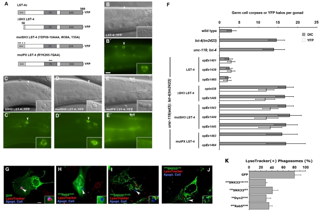
Clearance of apoptotic cells is of key importance during development, tissue homeostasis and wound healing in multi-cellular animals. Genetic studies in the nematode Caenorhabditis elegans have identified a set of genes involved in the early steps of cell clearance, in particular the recognition and internalization of apoptotic cells. A pathway that orchestrates the maturation of phagosomes containing ingested apoptotic cells in the worm has recently been described. However, many steps in this pathway remain elusive. Here we show that the C. elegans SNX9-family member LST-4 (lateral signaling target) and its closest mammalian orthologue SNX33 play an evolutionary conserved role during apoptotic cell corpse clearance. In lst-4 deficient worms, internalized apoptotic cells accumulated within non-acidified, DYN-1-positive but RAB-5-negative phagosomes. Genetically, we show that LST-4 functions at the same step as DYN-1 during corpse removal, upstream of the GTPase RAB-5. We further show that mammalian SNX33 rescue C. elegans lst-4 mutants and that overexpression of truncated SNX33 fragments interfered with phagosome maturation in a mammalian cell system. Taken together, our genetic and cell biological analyses suggest that LST-4 is recruited through a combined activity of DYN-1 and VPS-34 to the early phagosome membrane, where it cooperates with DYN-1 to promote recruitment/retention of RAB-5 on the early phagosomal membrane during cell corpse clearance. The functional conservation between LST-4 and SNX33 indicate that these early steps of apoptotic phagosome maturation are likely conserved through evolution.


Conflict of interest statement

Competing Interests: The authors have declared that no competing interests exist.

Figures

Figure 1
Figure 1. LST-4 is required for efficient cell corpse clearance in the adult C. elegans germ line.
(AD). DIC micrographs (AD) or epifluorescence pictures (A′D′) of C. elegans germlines. Dorsal is to the top and the germline bend is to right. Arrowheads indicate apoptotic germ cells or Acridine Orange (AO) staining of apoptotic corpses. In wild-type worms (A, D′) and in mutants with increased levels of germline apoptosis such as gla-3(op216) (B, B′), AO preferentially stains engulfed apoptotic cells present in acidic compartments. In worms mutant for genes required for efficient removal of apoptotic cells, here ced-12(k149), refractile cell corpses persist but do not stain with AO (C, C′). Similarly, lst-4(tm2423) worms show increased persistent cell corpses that fail to stain with AO (D, D′). Size bar, 10 µm. (EG′) Transmission electron microscopy images of cell corpses and their neighboring cells (EG) and corresponding camera lucida drawings (E′–G′); apoptotic cells are represented in dark grey, sheath cells in light grey and the germline syncytium in white. In lst-4(tm2423) animals (E, E′), apoptotic cells are fully internalized by the sheath cells, whereas in the ced-1(e1735); ced-5(n1812) double mutants apoptotic cells accumulate between germline syncytium and the sheath cells (F, F′). gla-3(op216) animals, which have increased germ cell apoptosis were used as a positive control (G, G′). Size bar, 2 µm. (H) Quantification of internalized apoptotic cells using TEM. For each genotype, two to three different animals 24 h post L4/adult molt were processed and analyzed as described by Zhou and coworkers . (I) The domain structure of the SNX9 subfamily of sorting nexins is conserved through evolution. The percent aminoacid identity of each domain between LST-4 and human SNX18, mouse SNX33 and fly DSH3PX1 is indicated. SH3: Src- homology 3 domain; PX: Phagocytic oxidase domain; BAR: Bin/Amphiphysin/Rvs domain. The tm2423 deletion results in a frame shift and a premature stop. (J) Expression of C. elegans LST-4 or mouse SNX33 in engulfing cells rescues the cell corpse clearance defect of lst-4 mutants. Results shown are mean ± s.d. n>15 animals for each genotype.
Figure 2
Figure 2. LST-4 is recruited to the early phagosome where it colocalizes with DYN-1 and RAB-5.
(AG) DIC micrographs (AD) or epifluorescence pictures (A′C′, EG) of C. elegans germ lines. Arrowheads indicate early apoptotic germ cells or protein localized around apoptotic germ cells. LST-4 is recruited around the apoptotic cell during internalization (A′, E, G arrowhead) and highlights early, SYTO negative corpses (F). SYTO, like Acridine Orange, preferentially stains late-stage, internalized apoptotic cells. When the internalization process is disrupted, as in ced-1(e1735) (B, B′) and ced-12(k149) mutants (C, C′), LST-4 localization around apoptotic germ cells is lost (B′, C′ arrowheads). (HO) DIC micrographs (H, L) or epifluorescence pictures (IK, MO) of C. elegans germ lines. LST-4::CFP (I, M) extensively colocalizes with DYN-1::YFP (J, K), but not with YFP::RAB-7 (N, O). (P) Localization of LST-4::YFP in different genetic backgrounds. Germ cell corpses and LST-4 positive phagosomes were quantified as described in materials and methods. Data shown are mean ± s.d. n>15 animals for each condition. Size bars, 10 µm. (R, S) Colocalization index of LST-4::CFP with YFP::actin, YFP::DYN-1, YFP::RAB-5 and YFP::RAB-7. Data shown are mean ± s.e.m. n>50 halos for each genotype. Determination of the colocalization index is described in materials and methods.
Figure 3
Figure 3. LST-4 function is required for DYN-1(+) to RAB-5(+) progression during phagosome maturation.
(AH′) DIC micrographs (AH) or epifluorescence pictures (A′H′) of wild type (AD′) or lst-4 mutant worms (EH′) carrying different fluorescent reporters. Arrowheads indicate early apoptotic germ cells or protein localized around apoptotic germ cells. Arrows denote late apoptotic germ cells or fluorescent halos formed around them. In both wild type (A′) and lst-4 mutants (E′) DYN-1::YFP is efficiently recruited to apoptotic cells whereas YFP::RAB-5 (B′, F′), YFP::2xFYVE (C′, G′) and YFP::RAB-7 (D′, H′) halos are less frequent in the gonad of lst-4(tm2423) mutant worms (F′, G′, H′ arrowheads) than in wild type animals (B′, C′, D′). (I) LST-4 is required for efficient recruitment of RAB-5, 2xFYVE and RAB-7 to the phagosome as well as for the release, but not for the recruitment of DYN-1. Results shown are mean ± s.d. n>15 animals for each genotype. (K) Genetic pathway for phagosome maturation. LST-4 likely acts downstream or in parallel to DYN-1 and upstream of RAB-5. Size bar, 10 mm.
Figure 4
Figure 4. Structure function analysis of LST-4/SNX33.
(AF) LST-4c constructs (A) were tested for their ability to rescue lst-4 mutants and for localization around internalized apoptotic cells (AD′). DIC (BE) and epifluorescence images (B′E′) of transgenic lines expressing LST-4c::YFP (B, B′), ΔSH3 LST-4c::YFP (C, C′), mutSH3 LST-4c::YFP (D, D′) and mutPX LST-4c::YFP (E, E′). (A) Schematic of LST-4c rescuing constructs. (F) Quantification of germ cell corpses and LST-4c::YFP halos around apoptotic cells in the corresponding genetic backgrounds. Animals were scored 24 h post L4/adult molt under DIC and epifluorescence. Data shown are means ± SD, n>15 animals. Size bar, 10 µm. (GK) The role of SNX33 in phagosome maturation is evolutionary conserved. NIH/3T3 fibroblasts transfected with GFP (G), GFPRab5S34N (H), YFPSNX33140–435 (I), YFPSNX33ΔPX (J), or HADyn2K44A were incubated with apoptotic thymocytes (shown in blue) and Lysotracker Red LTR to determine the efficiency of phagosome maturation. Cells transfected with YFPSNX33140–435, YFPSNX33ΔPX, GFPRab5S34N and HADyn2K44A showed decreased numbers of engulfed thymocytes (arrows) co-staining with Lysotracker Red. Size bar, 10 µm. The percentage internalized apoptotic phagosomes that were also Lysotracker positive (i.e., matured into acidic phagolysosomes) are shown in (K) (mean ± s.d., n = 3 experiments with 60 cells scored; only phagosomes in transfected [GFP(+) or HA(+)] cells were scored).
Figure 5
Figure 5. Model of early phagosome maturation.
1. DYN-1 is recruited to nascent phagosomes and subsequently recruits VPS-34. 2a. VPS-34 in turn promotes the recruitment of LST-4 through direct interaction and/or the production of phasphatidylinositol-3 phosphates (open arrow) to which LST-4 binds via its PX domain. 2b. Membrane bound LST-4 associates physically with DYN-1 to generate an active complex that promotes RAB-5 recruitment and further phagosome maturation. 3. The presence of active RAB-5 on the phagosome membrane stimulates the inactivation and release of the DYN-1/LST-4 complex from mature phagosomes.

References

    1. Henson PM, Hume DA. Apoptotic cell removal in development and tissue homeostasis. Trends Immunol. 2006;27:244–250. - PubMed
    1. Ravichandran KS, Lorenz U. Engulfment of apoptotic cells: signals for a good meal. Nat Rev Immunol. 2007;7:964–974. - PubMed
    1. Lettre G, Hengartner MO. Developmental apoptosis in C. elegans: a complex CEDnario. Nat Rev Mol Cell Biol. 2006;7:97–108. - PubMed
    1. Kinchen JM, Cabello J, Klingele D, Wong K, Feichtinger R, et al. Two pathways converge at CED-10 to mediate actin rearrangement and corpse removal in C. elegans. Nature. 2005;434:93–99. - PubMed
    1. Hurwitz ME, Vanderzalm PJ, Bloom L, Goldman J, Garriga G, et al. Abl kinase inhibits the engulfment of apoptotic [corrected] cells in Caenorhabditis elegans. PLoS Biol. 2009;7:e99. - PMC - PubMed

Publication types

MeSH terms

LinkOut - more resources