

The protective role of 5-hydroxymethyl-2-furfural (5-HMF) against acute hypobaric hypoxia
- PMID: 21494793
- PMCID: PMC3156263
- DOI: 10.1007/s12192-011-0264-8
The protective role of 5-hydroxymethyl-2-furfural (5-HMF) against acute hypobaric hypoxia
Abstract
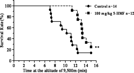
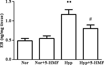
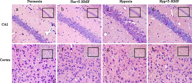
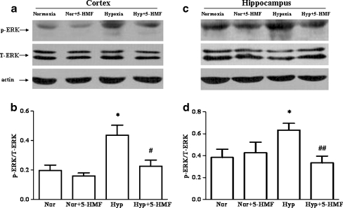
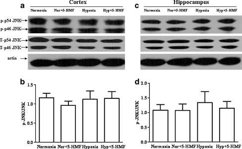
Our previous study showed that pretreatment with 5-hydroxymethyl-2-furfural (5-HMF) led to protection against hypoxic injury via a p-ERK-mediated pathway in vitro. Whether the protection of 5-HMF against hypoxia is effective in vivo is unknown. The present study is aimed to verify the role of 5-HMF in acute hypobaric hypoxia using Kunming mice as an in vivo model and further investigate the underlying mechanisms. Mice pretreated with or without 5-HMF for 1 h were exposed to acute hypobaric hypoxic condition for 6 h and then the survival time, the survival rate, the permeability of blood-brain barrier (BBB), the histological analysis in hippocampus and cortex, and the phosphorylation level of mitogen-activated protein kinases (ERK, JNK, and p38) were investigated. The results showed that 5-HMF significantly increased the survival time and the survival rate of mice. Accordingly, pretreatment with 5-HMF markedly attenuated acute hypobaric hypoxia-induced permeability of BBB (P < 0.01). In addition, the cellular damage extent of the hippocampus and the cortex induced by hypoxia for 6 h was also attenuated by pretreatment with 5-HMF, especially in the hippocampus CA1 region. Furthermore, the activation of ERK rather than JNK and p38 was involved in the protection of 5-HMF against acute hypobaric hypoxia. In summary, 5-HMF enhanced the survival capability of mice and decreased acute hypoxic damage to the brain, which may be associated with the effects on BBB and p-ERK.
Figures

Similar articles
-
The protective role of 5-HMF against hypoxic injury.Cell Stress Chaperones. 2011 May;16(3):267-73. doi: 10.1007/s12192-010-0238-2. Epub 2010 Nov 6. Cell Stress Chaperones. 2011. PMID: 21057989 Free PMC article.
-
Activation of p38 mitogen-activated protein kinase (p38 MAPK), extracellular signal-regulated kinase (ERK) and c-jun N-terminal kinase (JNK) during hypoxia in cerebral cortical nuclei of guinea pig fetus at term: role of nitric oxide.Neurosci Lett. 2008 Jul 4;439(1):94-9. doi: 10.1016/j.neulet.2008.02.037. Epub 2008 Feb 26. Neurosci Lett. 2008. PMID: 18511197
-
Hippocampal mitogen-activated protein kinase activation is associated with intermittent hypoxia in a rat model of obstructive sleep apnea syndrome.Mol Med Rep. 2016 Jan;13(1):137-45. doi: 10.3892/mmr.2015.4505. Epub 2015 Nov 5. Mol Med Rep. 2016. PMID: 26549199 Free PMC article.
-
The antisickling agent, 5-hydroxymethyl-2-furfural: Other potential pharmacological applications.Med Res Rev. 2024 Nov;44(6):2707-2729. doi: 10.1002/med.22062. Epub 2024 Jun 6. Med Res Rev. 2024. PMID: 38842004 Review.
-
Mitogen-Activated Protein Kinases and Hypoxic/Ischemic Nephropathy.Cell Physiol Biochem. 2016;39(3):1051-67. doi: 10.1159/000447812. Epub 2016 Aug 22. Cell Physiol Biochem. 2016. PMID: 27544204 Review.
Cited by
-
Several natural phytochemicals from Chinese traditional fermented food-pickled Raphanus sativus L.: Purification and characterization.Food Chem X. 2022 Jul 16;15:100390. doi: 10.1016/j.fochx.2022.100390. eCollection 2022 Oct 30. Food Chem X. 2022. PMID: 35874426 Free PMC article.
-
5-Hydroxymethylfurfural Ameliorates Allergic Inflammation in HMC-1 Cells by Inactivating NF-κB and MAPK Signaling Pathways.Biochem Genet. 2024 Jun;62(3):1521-1538. doi: 10.1007/s10528-023-10492-9. Epub 2023 Aug 30. Biochem Genet. 2024. PMID: 37648883
-
5-HMF attenuates inflammation and demyelination in experimental autoimmune encephalomyelitis mice by inhibiting the MIF-CD74 interaction.Acta Biochim Biophys Sin (Shanghai). 2023 Jul 10;55(8):1222-1233. doi: 10.3724/abbs.2023105. Acta Biochim Biophys Sin (Shanghai). 2023. PMID: 37431183 Free PMC article.
-
Effect of novel sequential soaking treatments on Maillard reaction products in potato and alternative vegetable crisps.Heliyon. 2021 Jun 29;7(7):e07441. doi: 10.1016/j.heliyon.2021.e07441. eCollection 2021 Jul. Heliyon. 2021. PMID: 34286122 Free PMC article.
-
A broad diversity in oxygen affinity to haemoglobin.Sci Rep. 2020 Oct 9;10(1):16920. doi: 10.1038/s41598-020-73560-9. Sci Rep. 2020. PMID: 33037242 Free PMC article.
References
-
- Beaumont A, Marmarou A, Hayasaki K, Barzo P, Fatouros P, Corwin F, Marmarou C, Dunbar J. The permissive nature of blood brain barrier (BBB) opening in edema formation following traumatic brain injury. Acta Neurochir Suppl. 2000;76:125–129. - PubMed
Publication types
MeSH terms
Substances
LinkOut - more resources
Full Text Sources
Other Literature Sources
Research Materials
Miscellaneous
