. 2011 May 23;50(22):5110-5.
doi: 10.1002/anie.201007824. Epub 2011 Apr 14.

Inhibition of amyloid peptide fibrillation by inorganic nanoparticles: functional similarities with proteins

Affiliations

Inhibition of amyloid peptide fibrillation by inorganic nanoparticles: functional similarities with proteins

Seong Il Yoo et al. Angew Chem Int Ed Engl. .

Abstract

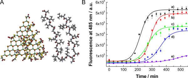
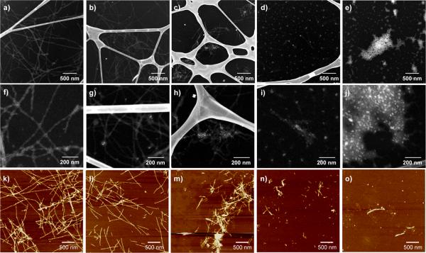
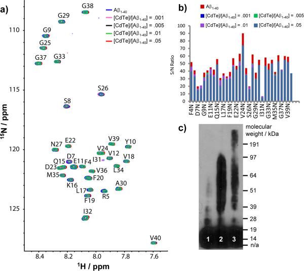
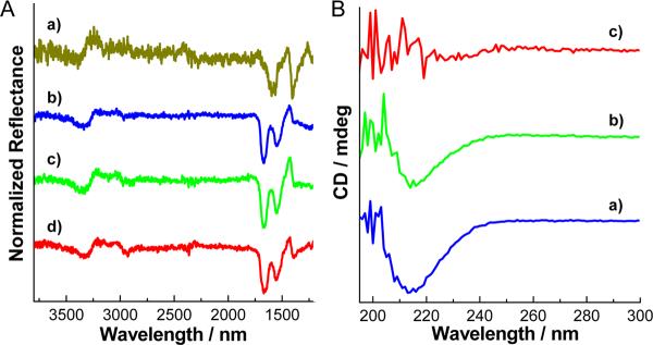
Aggregation of amyloid-β peptides (Aβ) into fibrils is the key pathological feature of many neurodegenerative disorders. Typical drugs inhibit Aβ fibrillation by binding to monomers in 1:1 ratio and display low efficacy. Here, we report that model CdTe nanoparticles (NPs) can efficiently prevent fibrillation of Aβ associating with 100–330 monomers at once. The inhibition is based on the binding multiple Aβ oligomers rather than individual monomers. The oligomer route of inhibition is associated with strong van der Waals interactions characteristic for NPs and presents substantial advantages in the mitigation of toxicity of the misfolded peptides. Molar efficiency and the inhibition mechanism revealed by NPs are analogous to those found for proteins responsible for prevention of amyloid fibrillation in human body. Besides providing a stimulus for finding biocompatible NPs with similar capabilities, these data suggest that inorganic NPs can mimic some sophisticated biological functionalities of proteins.


Figures

Figure 1
Figure 1
(A) Molecular structure of CdTe NPs and Aβ1-42 peptide. Some of the molecules of TGA adsorbed to the surface of CdTe are removed for clarity. Peptide is folded in the configuration characteristic for fibrils. Molecular modeling was carried out by SPARTAN. (B) Kinetics of Aβ1-40 fibrillation with and without CdTe NPs. Time-dependent ThT fluorescence was monitored at 485 nm with excitation wavelength of 450 nm. The molar ratio of [CdTe]/[Aβ1-40] was varied from 0 to 0.005: (a) 0, (b) 0.001, (c), 0.005, (d), 0.01 and (e) 0.05. Fluorescence intensity of ThT at 485 nm is proportional to the amount of fibrils.
Figure 2
Figure 2
TEM (a – j) and AFM (k – o) images of Aβ1-40 incubated with and without CdTe NPs. The molar ratios of [CdTe]/[Aβ1-40] were (a, f, k) 0, (b, g, l) 0.001, (c, h, m) 0.005, (d, i, n) 0.01, and (e, j, o) 0.05. In TEM images, NPs appeared as bright spots that can be easily distinguished from the fibrils even for small concentrations of NPs. The Z-contrast nature of the images enables the direct visualization of the peptide structures without a staining process.
Figure 3
Figure 3
NMR spectra (a, b) and Western blot (c) with and without CdTe NPs. (a) 2D SOFAST-HMQC spectra of freshly dissolved Aβ1-40 with increasing amounts of CdTe NPs at 10° C; (b) The change in the signal-to-noise ratio of Aβ1-40; (c) Western blot of freshly prepared Aβ1-42 (lane 1), 1 day incubated Aβ1-42 in the absence (lane 2) and presence (lane 3) of CdTe NPs. In Western blotting, the molar ratio of [CdTe]/[Aβ1-42] was 0.05. Notations for 1-letter abbreviation of each residue can be found in SI Text.
Figure 4
Figure 4
(A) FT-IR spectra of Aβ1-42 with and without CdTe NPs. (a) CdTe NPs, (b – d) Aβ1-42 with CdTe NPs. The molar ratios of [CdTe]/[Aβ1-42] were (b) 0, (c) 0.01, and (d) 0.05. (B) CD spectra of Aβ1-42 with and without CdTe NPs. The molar ratios of [CdTe]/[Aβ1-42] were (a) 0, (b) 0.01, and (c) 0.05. Both FT-IR and CD spectra were obtained after 1 day incubation. In Figure 4B trace c, CD signal below ~ 215 nm became noisy by the strong absorbance of CdTe NPs.

References

    1. Chiti F, Dobson CM. Annu. Rev. Biochem. 2006;75:333. - PubMed
    2. Lansbury PT, Lashuel HA. Nature. 2006;443:774. - PubMed
    3. Ross CA, Poirier MA. Nature Rev. Mol. Cell. Biol. 2005;6:891. - PubMed
    4. Hardy J, Selkoe DJ. Science. 2002;297:353. - PubMed
    1. Petkova AT, Leapman RD, Guo Z, Yau W-M, Mattson MP, Tycko R. Science. 2005;307:262. - PubMed
    2. Lorenzo A, Yankner BA. Proc. Natl. Acad. Sci. USA. 1994;91:12243. - PMC - PubMed
    1. Haass C, Selkoe DJ. Nature Rev. Mol. Cell. Biol. 2007;8:101. - PubMed
    2. Cleary JP, Walsh DM, Hofmeister JJ, Shankar GM, Kuskowski MA, Selkoe DJ, Ashe KH. Nature Neurosci. 2004;8:79. - PubMed
    3. Bitan G, Kirkitadze MD, Lomakin A, Vollers SS, Benedek GB, Teplow DB. Proc. Natl. Acad. Sci. USA. 2003;100:330. - PMC - PubMed
    4. Walsh DM, Klyubin I, Fadeeva JV, Cullen WK, Anwyl R, Wolfe MS, Rowan MJ, Selkoe DJ. Nature. 2002;416:535. - PubMed
    5. Klein WL, Krafft GA, Finch CE. Trends Neurosci. 2001;24:219. - PubMed
    1. Takahashi T, Mihara H. Accounts Chem. Res. 2008;41:1309. - PubMed
    1. Gordon DJ, Sciarretta KL, Meredith SC. Biochemistry. 2001;40:8237. - PubMed
    2. Lowe TL, Strzelec A, Kiessling LL, Murphy RM. Biochemistry. 2001;40:7882. - PubMed
    3. Findeis MA, Musso GM, Arico-Muendel CC, Benjamin HW, Hundal AM, Lee J-J, Chin J, Kelley M, Wakefield J, Hayward NJ, Molineaux SM. Biochemistry. 1999;38:6791. - PubMed
    4. Tjernberg LO, Näslund J, Lindqvist F, Johansson J, Karlström AR, Thyberg J, Terenius L, Nordstedt C. J. Biol. Chem. 1996;271:8545. - PubMed
    5. Soto C, Kindy MS, Baumann M, Frangione B. Biochem. Biophys. Res. Commun. 1996;226:672. - PubMed

Publication types

MeSH terms