. 2011 Jun 27;50(27):6109-14.
doi: 10.1002/anie.201100884. Epub 2011 Apr 14.

Tunable, ultrasensitive pH-responsive nanoparticles targeting specific endocytic organelles in living cells

Affiliations

Tunable, ultrasensitive pH-responsive nanoparticles targeting specific endocytic organelles in living cells

Kejin Zhou et al. Angew Chem Int Ed Engl. .
No abstract available


Figures

Figure 1
Figure 1
a) Schematic design of pH-activatable micelle (pHAM) nanoprobes. At pH > pKa of ammonium groups (left panel), the neutralized PR segments self-assemble into the micelle cores, leading to quenching of fluorophores due to homoFRET and PeT mechanisms. Upon pH activation (pH < pKa, right panel), formation of charged ammonium groups results in micelle dissociation into unimers with dramatic increase in fluorescence emission. b) Structures of the PEO-b-(PR-r-TMR) copolymers in the di-alkyl and cyclic series.
Figure 2
Figure 2
Illustration of tunable, ultra-pH responsive properties of pHAM nanoprobes. a) Representative fluorescent images of different nanoprobe solutions (6, 3, 4) at the same polymer concentration (0.1 mg/mL) but different pH values. A narrow pH response is observed for each nanoprobe at different transition pHs. Copolymer 1 serves as an always “ON” control without pH response. A blue light (λex = 440 ~ 480 nm, 450 mW/cm2) was used to excite the nanoprobes. b) Normalized fluorescence intensity as a function of pH for different pHAM nanoprobes. The pH response (ΔpH10–90%) is <0.25 pH unit and Fmax/Fmin is up to 55-fold (Supplementary Table S2). c) Stopped flow fluorescence measurement of nanoprobe 4 (pHt = 5.4) after pH activation at 4.9. Fluorescence recovery time (τ1/2) is 3.7 ms. Other pHAM nanoprobes show similarly fast kinetics (Table S2).
Figure 3
Figure 3
Investigation of the ultra-pH responsive properties of a representative pHAM. a) pH titration curves of copolymers 5 and 7 and their corresponding monomers. The added volumes of NaOH (VNaOH) were normalized to the initial amount of amine residues ([R3N]0) in mmol. b) 1H NMR spectra (in D2O) of 5 and 7 at different ionization states of the copolymers. c) TEM of 7 in pH 5.5 and 7.4 buffers at the polymer concentration of 2 mg/mL.
Figure 4
Figure 4
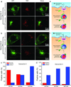
Investigation of subcellular activation of nanoprobes 3 and 4 in different endocytic organelles in human H2009 cells. a, b) Representative confocal images of activated nanoprobe 3 (a, pHt = 6.3) and 4 (b, pHt = 5.4) in cells with GFP-labeled early endosomes (top panel) and late endosomes/lysosomes (bottom panel) at 30 and 45 min, respectively. c, d) Percentage of positive cells (N=30–50 cells) with activated nanoprobe 3 and 4 colocalizing with early endosomes or late endosomes/lysosomes at different incubation times, respectively. e, f) Schematic illustration of the selective activations of nanoprobe 3 in early endosomes (pH 5.9–6.2) and 4 in late endosomes/lysosomes (pH 5.0–5.5), respectively.
Figure 5
Figure 5
Inhibition of lysosomal acidification by bafilomycin A1 and its effect on intracellular activation of nanoprobe 3 in H2009 cells. a) Confocal images of cells treated with bafilomycin A1 for 1 h followed by nanoprobe 3 incubation for 1 h. Lack of activation of nanoprobe 3 was observed as demonstrated by the absence of TMR fluorescence. b) Confocal images of the same H2009 cells after removal of bafilomycin A1 and nanoprobe 3 and incubation for additional 5 h. Nanoprobe activation was observed as indicated by the red fluorescence inside lysosomes (yellow dots in the overlay images). Scale bar = 20 µm.

References

    1. Singhal S, Nie S, Wang MD. Annu. Rev. Med. 2010;61:359–373. - PMC - PubMed
    2. Riehemann K, Schneider SW, Luger TA, Godin B, Ferrari M, Fuchs H. Angew. Chem. Int. Ed. 2009;48:872–897. - PMC - PubMed
    3. Weissleder R, Pittet MJ. Nature. 2008;452:580–589. - PMC - PubMed
    4. Davis ME, Chen Z, Shin DM. Nat. Rev. Drug Discov. 2008;7:771–782. - PubMed
    5. Peer D, Karp JM, Hong S, Farokhzad OC, Margalit R, Langer R. Nat. Nanotech. 2007;2:751–760. - PubMed
    1. Torchilin V. Eur. J. Pharm. Biopharm. 2009;71:431–444. - PMC - PubMed
    2. Lee ES, Gao ZG, Bae YH. J. Controlled Release. 2008;132:164–170. - PMC - PubMed
    1. Lee ES, Na K, Bae YH. J. Controlled Release. 2003;91:103–113. - PubMed
    2. Bae Y, Fukushima S, Harada A, Kataoka K. Angew. Chem. Int. Ed. 2003;42:4640–4643. - PubMed
    3. Lynn DM, Amiji MM, Langer R. Angew. Chem. Int. Ed. 2001;40:1707–1710. - PubMed
    1. Choi SW, Zhang Y, Xia Y. Angew. Chem. Int. Ed. 2010;49:7904–7908. - PMC - PubMed
    2. Jeong B, Bae YH, Kim SW. J. Controlled Release. 2000;63:155–163. - PubMed
    1. Wang C, Chen Q, Wang Z, Zhang X. Angew. Chem. Int. Ed. 2010;49:8612–8615. - PubMed
    2. Olson ES, Jiang T, Aguilera TA, Nguyen QT, Ellies LG, Scadeng M, Tsien RY. Proc. Natl. Acad. Sci. USA. 2010;107:4311–4316. - PMC - PubMed
    3. Bernardos A, Aznar E, Marcos MD, Martínez-Máñez R, Sancenón F, Soto J, Barat JM, Amorós P. Angew. Chem. Int. Ed. 2009;48:5884–5887. - PubMed

Publication types

LinkOut - more resources