Review
. 2011:80:375-401.
doi: 10.1146/annurev-biochem-060409-093259.

Reporting from the field: genetically encoded fluorescent reporters uncover signaling dynamics in living biological systems

Affiliations
Review

Reporting from the field: genetically encoded fluorescent reporters uncover signaling dynamics in living biological systems

Sohum Mehta et al. Annu Rev Biochem. 2011.

Abstract

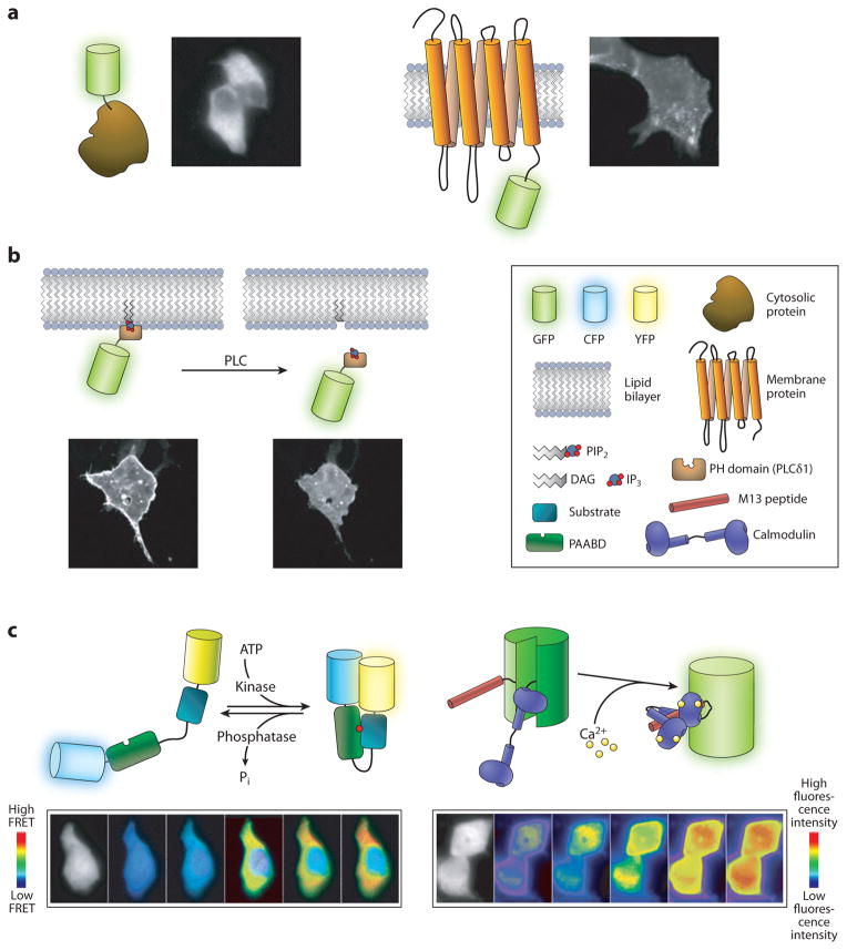
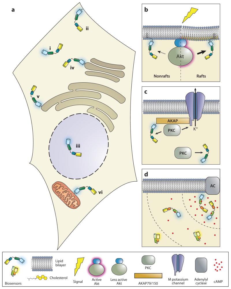
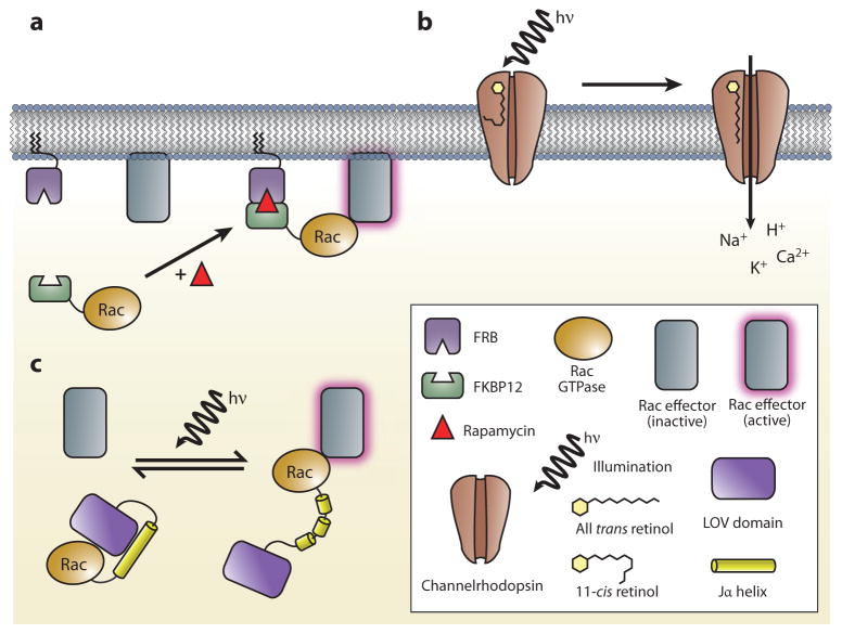
Real-time visualization of a wide range of biochemical processes in living systems is being made possible through the development and application of genetically encoded fluorescent reporters. These versatile biosensors have proven themselves tailor-made to the study of signal transduction, and in this review, we discuss some of the unique insights that they continue to provide regarding the spatial organization and dynamic regulation of intracellular signaling networks. In addition, we explore the more recent push to expand the scope of biological phenomena that can be monitored using these reporters, while also considering the potential to integrate this highly adaptable technology with a number of emerging techniques that may significantly broaden our view of how networks of biochemical processes shape larger biological phenomena.


Figures

Figure 1
Figure 1
Major classes of genetically encoded fluorescent reporters. (a) Reporters for visualizing the native localization of endogenous proteins. Direct fusion to a fluorescent protein enables observation of the subcellar distribution of numerous proteins. Shown are epifluorescence images of MIN6 cells expressing a fluorescent protein-tagged form of the type II regulatory subunit of protein kinase A (PKA) (left) and HEK293 cells expressing the β1 adrenergic receptor tagged with a fluorescent protein (right), along with respective schematic illustrations of green fluorescent protein (GFP) fused to either a cytosolic (left) or a transmembrane protein (right). (b) Reporters based on effector-guided translocation. Fusion of a fluorescent protein to a specific effector domain can be used to monitor second messenger dynamics. As illustrated here, conversion of phosphatidylinositol (4,5)-bisphosphate (PIP2) into inositol (1,4,5)-trisphosphate (IP3) and diacylglycerol (DAG) causes dissociation of a particular fluorescent protein-tagged pleckstrin homology (PH) domain from the plasma membrane. Confocal fluorescence images show reporter distribution before (left) and after (right) phospholipase C (PLC) activation in Cos-7 cells. (c) Reporters in which a molecular switch is used to detect a biochemical event. Molecular switches can be designed to respond to a variety of parameters. Fluorescence resonance energy transfer (FRET)-based reporters utilize molecular switches sandwiched between donor and acceptor fluorescent proteins, such as CFP and YFP (left). In a kinase activity reporter, phosphorylation (red circle) mediates conformational changes in the kinase activity-dependent molecular switch, modulating FRET between the fluorescent protein pair. Alternatively, a molecular switch based on a pair of interacting proteins, such as calmodulin (CaM) and the M13 peptide, can be inserted within a fluorescent protein (right). Ca2+ binding causes a conformational rearrangement that increases fluorescence intensity. Images in panel c depict HeLa cells expressing the PKA activity reporter AKAR, along with ratiometric images showing the FRET response of AKAR upon forskolin treatment (left, bottom), and HEK293 cells expressing the Ca2+ indicator GCaMP responding to thapsigargin treatment (right, bottom). Images colored to reflect increasing FRET (left) or increasing fluorescence intensity (right).
Figure 2
Figure 2
Studying compartmentalized signaling with subcellularly targeted biosensors. (a) Local signaling within and around subcellular organelles. By incorporating a particular targeting motif into the primary sequence of a genetically encoded reporter, it is possible to detect specific signaling activities at subcellular loci including, but not limited to (i) the cytosol, (ii) the plasma membrane, (iii) the nucleus, (iv) the Golgi apparatus, (v) the endoplasmic reticulum, and (vi) the mitochondria. (b) Signaling events within membrane microdomains. Reporters can be further localized to membrane microdomains using specific targeting motifs and can distinguish between raft- and nonraft-associated signaling activities. (c) Coordinated macromolecular signalosomes. Instead of relying on subcellular targeting motifs, reporters can be targeted to macromolecular complexes, for example, via direct fusion to scaffolding proteins, allowing for visualization of biochemical events specific to these complexes. (d ) Diffusible biochemical gradients. Biosensors can also be used to observe gradients of signaling activity. Given that it can rapidly respond to changes in its immediate environment, the response of a reporter will vary depending on its position within the gradient. AC, adenylyl cyclase; AKAP, A-kinase anchoring protein; Akt, a serine-threonine protein kinase; PKC, protein kinase C.
Figure 3
Figure 3
Three examples of targeted perturbations. (a) In chemically inducible dimer formation, rapamycin drives the association of an FKBP-rapamycin-binding domain (FRB domain) with the 12-kDa FK506-binding protein (FKBP12). This can be used, for example, to direct the activity of a given enzyme toward substrates in a location-specific manner. (b) Channelrhodopsins are relatives of the bacterial rhodopsins. These ion channels are inactive in the dark and become activated upon the light-induced isomerization of an associated retinol moiety. (c) Photoactivatable Rac consists of the light-oxygen-voltage (LOV) domain and the Jαhelical extension from the Avena sativa phototropin 1 protein fused to the N terminus of a constitutively active Rac isoform. In the absence of illumination, a complex between the LOV and Jαdomains prevents Rac from interacting with its effector proteins. A reversible, light-induced conformational change disrupts the LOV-Jα complex, permitting the expression of Rac activity with a high degree of temporal and spatial control.

Similar articles

Cited by

References

    1. Coons AH, Creech HJ, Jones RN, Berliner E. The demonstration of pneumococcal antigen in tissues by the use of fluorescent antibody. J Immunol. 1942;45:159–70.
    1. Bloom FE, Wedner HJ, Parker CW. The use of antibodies to study cell structure and metabolism. Pharmacol Rev. 1973;25:343–58. - PubMed
    1. Heffetz D, Fridkin M, Zick Y. Antibodies directed against phosphothreonine residues as potent tools for studying protein phosphorylation. Eur J Biochem. 1989;182:343–48. - PubMed
    1. Kaufmann H, Bailey JE, Fussenegger M. Use of antibodies for detection of phosphorylated proteins separated by two-dimensional gel electrophoresis. Proteomics. 2001;1:194–99. - PubMed
    1. Ross AH, Baltimore D, Eisen HN. Phosphotyrosine-containing proteins isolated by affinity chromatography with antibodies to a synthetic hapten. Nature. 1981;294:654–56. - PubMed

Publication types

MeSH terms

LinkOut - more resources