. 2011 Jul;219(1):44-52.
doi: 10.1111/j.1469-7580.2010.01330.x. Epub 2010 Dec 24.

Astrocyte phenotypes and their relationship to myelination

Affiliations

Astrocyte phenotypes and their relationship to myelination

Besma Nash et al. J Anat. 2011 Jul.

Abstract

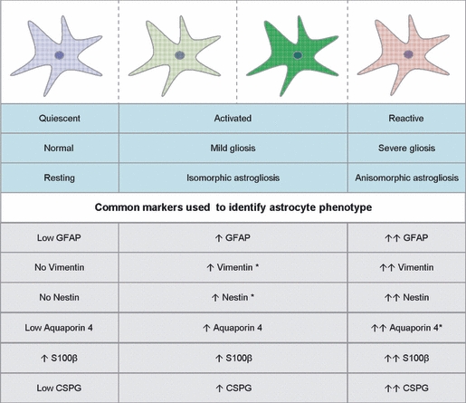
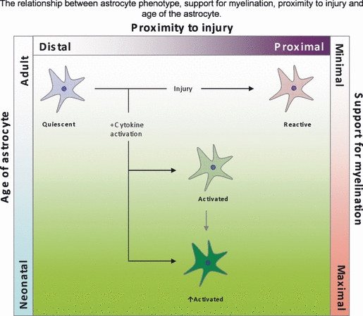
Astrocytes are one of the major glial cell types that maintain homeostasis in the undamaged CNS. After injury and disease, astrocytes become reactive and prevent regeneration; however, it has also been suggested that astrocytes can become activated and promote regeneration. Thus, it is hypothesised that astrocytes have an important role in modulating CNS repair. This review will focus on the variable phenotypic state of astrocytes that range from inactive/quiescent to reactive, and relate these to their ability to influence myelination. Using myelinating cultures plated on astrocytes we propose a possible mechanism for oligodendrocyte precursor cell interaction with the axon, leading to myelination. The phenotypic status of astrocytes is an intriguing and widely discussed issue, which is critical for understanding the mechanisms involved in CNS injury and its subsequent repair.


Figures

Fig. 1
Fig. 1
Stages of myelination visualised from the myelinating cultures immunolabelled with SMI-31 to depict neurites (red) and PLP (green) for mature myelin. Stages of myelination in vitro can be followed in the cultures. (A) At early stages of myelination, OPCs wrap a myelin sheath as a spiral around the axons (arrow). Image taken after 18 days in culture. (B) The myelin sheath begins to fill in around from the initial spirals around the axon. Image taken at 18 days in culture. (C) Oligodendrocytes can make many internodes of myelin often from primary processes (*), but sometimes from secondary (**). (D) The myelinating cultures form many nodes of Ranvier. Image taken at 22–28 days in culture.
Fig. 2
Fig. 2
Schematic for a mechanism for myelination based on images obtained from the myelinating cultures. (A) The glial cell puts out many processes and spirals a sheath around the axon. (B) The spiral of myelin fills forming a larger expanse of membrane. (C) Eventually this fills out to form myelin internodes. (Ai) OPCs wrapping a spiral of myelin around the axon. (Aii) An example of an oligodendrocyte extending multiple processes towards axons. (Bi) The myelin from the membrane spiral expands out along the axon. (Ci) Oligodendrocytes form internodes of myelin separated by nodal spacing over several axons. Myelinating cultures were immunolabelled with SMI-31 to depict neurites (red) and PLP (green) for mature myelin.
Fig. 3
Fig. 3
Summary of astrocyte phenotypes and markers used to identify them. The astrocyte can be termed quiescent in normal adult resting CNS tissue. They can become activated by various mechanisms that result in mild/isomorphic astrogliosis, with a mild increase in cytoskeleton markers. Activated astrocytes are also more distal to the injury site. The most severe phenotype is the reactive/anisomorphic astrocyte that forms at the gliotic scar. This phenotype is characterised by a high increase in cytoskeletal molecules (Ridet et al. 1997; Fawcett & Asher, 1999Liberto et al. 2004; East et al. 2009; Sofroniew & Vinters, 2010). Asterisks represent our predicted information on expression of these markers. Arrows indicate direction of expression and intensity. CSPG, chondroitin sulphate proteoglycans; GFAP, glial fibrillary acidic protein.
Fig. 4
Fig. 4
Summary of how an astrocyte phenotype, which is determined by age and location, can influence myelination. Astrocytes in close proximity to the site of injury are typically described as reactive and their support for myelination is poor. Astrocytes more distal to the site of injury receive ‘injury’ signal in the form of cytokines and increase their state of activation, ultimately increasing their support for oligodendrocytes via the secretion of trophic factors. Astrocytes in the neonatal animals respond better to injury possibly due to the reactivation state of the astrocytes, and are therefore more equipped to support myelination than in the adult. Green represents a more positive effect on myelination, while red represents a more negative effect on myelination.

Similar articles

Cited by

References

    1. Albrecht PJ, Dahl JP, Stoltzfus OK, et al. Ciliary neurotrophic factor activates spinal cord astrocytes, stimulating their production and release of fibroblast growth factor-2, to increase motor neuron survival. Exp Neurol. 2002;173:46–62. - PubMed
    1. Alexander WS. Progressive fibrinoid degeneration of fibrillary astrocytes associated with menal retardation in a hydrocephalic infant. Brain. 1949;72:373–381. - PubMed
    1. Back SA, Tuohy TM, Chen H, et al. Hyaluronan accumulates in demyelinated lesions and inhibits oligodendrocyte progenitor maturation. Nat Med. 2005;11:966–972. - PubMed
    1. Barrett CP, Donati EG, Guth L. Differences between adult and neonatal rats in their astroglial response to spinal injury. Exp Neurol. 1984;84:374–385. - PubMed
    1. Bauer NG, Richter-Landsberg C, ffrench-Constant C. Role of the oligodendrocyte cytoskeleton in differentiation and myelination. Glia. 2009;57:1790–1801. - PubMed

Publication types

LinkOut - more resources