

Neuroprotective properties of marrow-isolated adult multilineage-inducible cells in rat hippocampus following global cerebral ischemia are enhanced when complexed to biomimetic microcarriers
- PMID: 21496021
- PMCID: PMC4516086
- DOI: 10.1111/j.1471-4159.2011.07272.x
Neuroprotective properties of marrow-isolated adult multilineage-inducible cells in rat hippocampus following global cerebral ischemia are enhanced when complexed to biomimetic microcarriers
Abstract
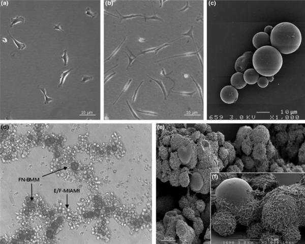
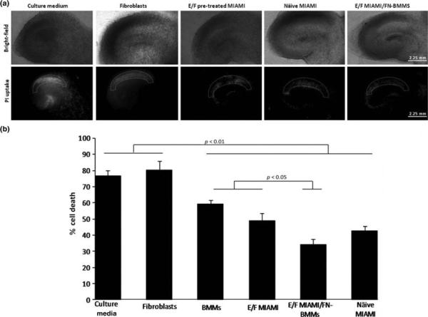
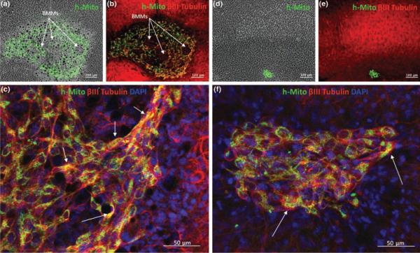
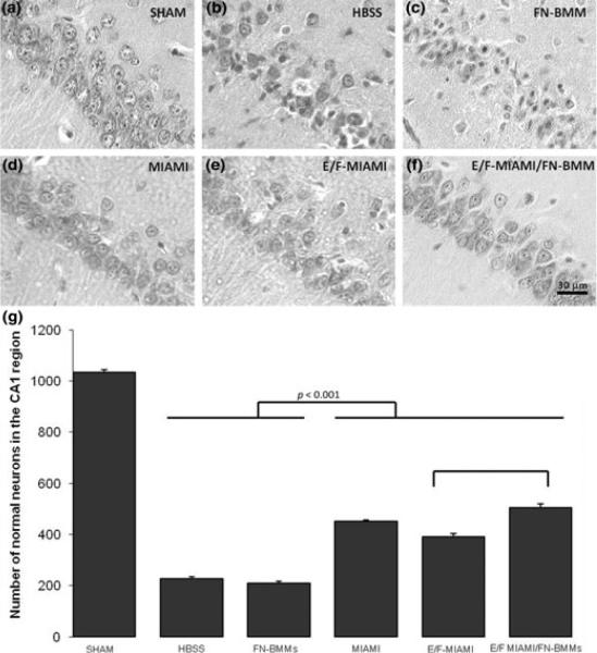
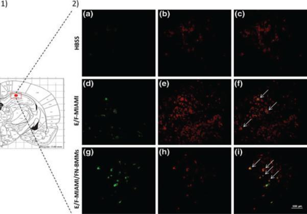
Cell-based therapies for global cerebral ischemia represent promising approaches for neuronal damage prevention and tissue repair promotion. We examined the potential of marrow-isolated adult multilineage-inducible (MIAMI) cells, a homogeneous subpopulation of immature human mesenchymal stromal cell, injected into the hippocampus to prevent neuronal damage induced by global ischemia using rat organotypic hippocampal slices exposed to oxygen-glucose deprivation and rats subjected to asphyxial cardiac arrest. We next examined the value of combining fibronectin-coated biomimetic microcarriers (FN-BMMs) with epidermal growth factor (EGF)/basic fibroblast growth factor (bFGF) pre-treated MIAMI compared to EGF/bFGF pre-treated MIAMI cells alone, for their in vitro and in vivo neuroprotective capacity. Naïve and EGF/bFGF pre-treated MIAMI cells significantly protected the Cornu Ammonis layer 1 (CA1) against ischemic death in hippocampal slices and increased CA1 survival in rats. MIAMI cells therapeutic value was significantly increased when delivering the cells complexed with FN-BMMs, probably by increasing stem cell survival and paracrine secretion of pro-survival and/or anti-inflammatory molecules as concluded from survival, differentiation and gene expression analysis. Four days after oxygen and glucose deprivation and asphyxial cardiac arrest, few transplanted cells administered alone survived in the brain whereas stem cell survival improved when injected complexed with FN-BMMs. Interestingly, a large fraction of the transplanted cells administered alone or in complexes expressed βIII-tubulin suggesting that partial neuronal transdifferentiation may be a contributing factor to the neuroprotective mechanism of MIAMI cells.
© 2011 The Authors. Journal of Neurochemistry © 2011 International Society for Neurochemistry.
Figures
Similar articles
-
The therapeutic potential of human multipotent mesenchymal stromal cells combined with pharmacologically active microcarriers transplanted in hemi-parkinsonian rats.Biomaterials. 2011 Feb;32(6):1560-73. doi: 10.1016/j.biomaterials.2010.10.041. Epub 2010 Nov 12. Biomaterials. 2011. PMID: 21074844
-
Survival, differentiation, and neuroprotective mechanisms of human stem cells complexed with neurotrophin-3-releasing pharmacologically active microcarriers in an ex vivo model of Parkinson's disease.Stem Cells Transl Med. 2015 Jun;4(6):670-84. doi: 10.5966/sctm.2014-0139. Epub 2015 Apr 29. Stem Cells Transl Med. 2015. PMID: 25925835 Free PMC article.
-
Systemic administration of mesenchymal stem cells increases neuron survival after global cerebral ischemia in vivo (2VO).Neural Plast. 2010;2010:534925. doi: 10.1155/2010/534925. Epub 2010 Dec 19. Neural Plast. 2010. PMID: 21331297 Free PMC article.
-
Pharmacologically active microcarriers delivering BDNF within a hydrogel: Novel strategy for human bone marrow-derived stem cells neural/neuronal differentiation guidance and therapeutic secretome enhancement.Acta Biomater. 2017 Feb;49:167-180. doi: 10.1016/j.actbio.2016.11.030. Epub 2016 Nov 16. Acta Biomater. 2017. PMID: 27865962
-
Organotypic cultures as tools for optimizing central nervous system cell therapies.Exp Neurol. 2013 Oct;248:429-40. doi: 10.1016/j.expneurol.2013.07.012. Epub 2013 Jul 27. Exp Neurol. 2013. PMID: 23899655 Review.
Cited by
-
Characterization of three washing/decellularization procedures for the production of bioactive human micronized neural tissue (hMINT).Cell Tissue Bank. 2023 Dec;24(4):693-703. doi: 10.1007/s10561-023-10075-3. Epub 2023 Feb 28. Cell Tissue Bank. 2023. PMID: 36854877
-
Marrow-isolated adult multilineage inducible cells embedded within a biologically-inspired construct promote recovery in a mouse model of peripheral vascular disease.Biomed Mater. 2017 Feb 17;12(1):015024. doi: 10.1088/1748-605X/aa5a74. Biomed Mater. 2017. PMID: 28211362 Free PMC article.
-
Superparamagnetic Iron Oxide Nanoparticles in Musculoskeletal Biology.Tissue Eng Part B Rev. 2017 Aug;23(4):373-385. doi: 10.1089/ten.TEB.2016.0437. Epub 2017 Jan 11. Tissue Eng Part B Rev. 2017. PMID: 27998240 Free PMC article.
-
Platelet-rich plasma promotes the proliferation of human muscle derived progenitor cells and maintains their stemness.PLoS One. 2013 Jun 7;8(6):e64923. doi: 10.1371/journal.pone.0064923. Print 2013. PLoS One. 2013. PMID: 23762264 Free PMC article.
-
Bone marrow mesenchymal stem cell therapy in ischemic stroke: mechanisms of action and treatment optimization strategies.Neural Regen Res. 2016 Jun;11(6):1015-24. doi: 10.4103/1673-5374.184506. Neural Regen Res. 2016. PMID: 27482235 Free PMC article. Review.
References
-
- Bang OY, Lee JS, Lee PH, Lee G. Autologous mesenchymal stem cell transplantation in stroke patients. Ann Neurol. 2005;57:874–882. - PubMed
-
- Bible E, Chau DY, Alexander MR, Price J, Shakesheff KM, Modo M. The support of neural stem cells transplanted into stroke-induced brain cavities by PLGA particles. Biomaterials. 2009;30:2985–2994. - PubMed
-
- Caplan AI, Dennis JE. Mesenchymal stem cells as trophic mediators. J Cell Biochem. 2006;98:1076–1084. - PubMed
Publication types
MeSH terms
Substances
Grants and funding
LinkOut - more resources
Full Text Sources
Other Literature Sources
Miscellaneous
